

서론
한국에서 딸기 재배면적은 6,890 ha이고, 연간 생산량은 316,803톤이다(Ministry of Agriculture, Food and Rural Affairs, 2014). 딸기세균병은 1962년 미국 미네소타에서 Xanthomonas fragariae에 의한 딸기세균모무늬병(bacterial angular leaf spot of strawberry) (Kennedy와 King, 1962a)과 이탈리아 체제나(Cesena)에서 X. arboricola pv. fragariae에 의한 딸기세균잎마름병(bacterial leaf blight of strawberry) (Janse 등, 2001)이 보고되었다. 한국에서는 2010년 7월 경상남도 진주시 수곡면과 하동군 옥동면에서 X. fragariae에 의한 딸기세균모무늬병 발생이 보고되었다(Kwon 등, 2010).
딸기세균모무늬병은 딸기생산에 중요한 병이다. 본 병에 의한 경제적 손실은 정확히 기록되어 있지 않으나 미국 플로리다에서 연간 8% (Roberts 등, 1997), 위스콘신에서 75% (Epstein, 1996), 유럽에서 10%-20% (Elphinston, 2005) 생산량을 감소시키는 것으로 알려져 있다. 또한, 꽃받침 감염에 의하여 상품성을 저하시킨다(Maas 등, 1995). 그리고 엽병과 지제부에 병이 발생하면 전신 감염되어 묘가 죽게된다(Hildebrand 등, 1967). X. fragariae는 유럽지중해식물보호기구에 A2 검역병원균으로 등재되어 있고(European and Mediterranean Plant Protection Organization, 2010), 국내에도 국가 검역대상으로 등록되어 있다(National Plant Quarantine Service, 2008). 특히, 유럽으로 재식용 딸기를 수출하려면 고강도의 식물검역과정을 거쳐야 한다.
본 병의 국내 발생보고(Kwon 등, 2010)로 2012년 농촌진흥청 외래병해충 방제대책회의에서 딸기세균모무늬병의 공적방제 필요성이 결정되었다. 이후 병 확산을 억제하기 위하여 2013년 3월부터 병 발생 지역과 인근 지역의 딸기재배 농가를 대상으로 딸기세균모무늬병과 병 발생 억제 방법에 관한 교육 및 홍보를 시작하였다. 딸기세균모무늬병의 화학적 방제는 어렵다. 스트렙토마이신과 옥시테트라사이클린 같은 항생제는 병원세균 자체에 대해 효과적(Alippi 등, 1989)이나 포장에서 사용할 때 약제 저항성균이 출현될 수 있고(Stall과 Thayer, 1962), 현재까지 딸기세균모무늬병균 방제용 등록된 항생제가 없다(Roberts 등, 1997). 딸기세균모무늬병은 건전한 모종 사용, 재배지 습도와 양분관리, 보호살균제 살포 등의 방법으로 예방하고 있다. 병이 발생되면 동제 화합물과 만코제브 혼합물 살포로 병을 억제시킬 수 있으나 이들 화합물의 잦은 살포는 식물에 독성을 끼칠 수 있다(Conover와 Gerhold, 1981; Marco와 Stall, 1983; Roberts 등, 1997). 그러므로 저항성 품종 재배가 가장 효과적인 딸기세균모무늬병 방제수단이다. 그러나 현재 국내에는 딸기세균모무늬병에 대한 재배품종 저항성 조사가 없다.
본 연구에서는 딸기세균모무늬병 공적 방제를 위한 병 발생 지역을 확인하고, 병 발생 지역에서 경시적 병 발생을 조사하였다. 그리고 국내에서 분리된 딸기세균모무늬병균의 생리생화학적, 유전학적 특성과 국내 재배되고 있는 딸기품종들의 저항성 특성 등을 조사하였다.
재료 및 방법
딸기세균모무늬병 발생조사
2012년 11월 충청남도와 제주도를 제외한 전국 1,482개 딸기재배농가에서 딸기세균모무늬병 발생을 조사하였다. 또한 2012년 1월부터 2015년 1월까지 딸기세균모무늬병이 처음 발견된 진주시 수곡면과 하동군 옥동면 딸기재배포장에서 딸기세균모무늬병 발생 변화를 관찰하였다. 수곡면과 하동군의 딸기재배포장을 임의로 각 40포장을 선정하여 각 재배포장에서 병 발생을 조사하였다.
병원균분리
2011년 10월 경남 진주시 수곡면과 하동군 옥종면에서 딸기세균모무늬병 증상이 있는 병든 조직을 수거하여 실험실에서 병원세균을 분리하였다. 딸기세균모무늬병징이 있는 조직을 70% 알코올에 30초 동안 표면살균하였다. 표면살균된 병든 부위를 작은 조각으로 자르고 50 μl 살균수에 담근 후 살균된 루프로 마쇄하였다. 마쇄된 현탁액을 살균된 플라스틱 루프에 묻혀 yeast extract-dextrose-CaCO (YDC) 한천 배양기에 획선배양하였다. 접종된 배양기를 26°C에서 4-7일 배양하여 병원세균을 분리하였다. 순수 분리된 병원균은 20% glycerol 용액에 혼합 후 70°C 보관하여 필요할 때 사용하였다(Table 1).
Table 1
Strains of Xanthomonas fragariae used in this study, and GenBank accession numbers
| Bacterial name (strain source*) | Code No. | Location | Isolation year | GenBank accession number | Reference | |||
|---|---|---|---|---|---|---|---|---|
|
|
||||||||
| dnaK | fyuA | gyrB | rpoD | |||||
| X. fragariae (LMG 708T) | BC 2719 | USA | 1960 | KT886342 | KT886357 | KT886372 | KT886381 | This study |
| X. fragariae | BC 3161 | Okjong, Hadong | 2010 | KT886343 | KT886358 | KT886373 | KT886382 | This study |
| X. fragariae | BC 3162 | Okjong, Hadong | 2010 | KT886344 | KT886359 | KT886374 | KT886383 | This study |
| X. fragariae | BC 3163 | Okjong, Hadong | 2010 | NT | NT | NT | NT | This study |
| X. fragariae | BC 3164 | Okjong, Hadong | 2010 | NT | NT | NT | NT | This study |
| X. fragariae | BC 3165 | Sukok, Jinju | 2010 | KT886345 | KT886360 | KT886375 | KT886384 | This study |
| X. fragariae | BC 3166 | Sukok, Jinju | 2010 | KT886346 | KT886361 | KT886376 | KT886385 | This study |
| X. fragariae | BC 3167 | Sukok, Jinju | 2010 | KT886347 | KT886362 | KT886377 | KT886386 | This study |
| X. fragariae | BC 3168 | Sukok, Jinju | 2010 | NT | NT | NT | NT | This study |
| X. fragariae | BC 3169 | Sukok, Jinju | 2010 | KT886348 | KT886363 | KT886378 | KT886387 | This study |
| X. fragariae | BC 3181 | Sukok, Jinju | 2010 | NT | NT | NT | NT | This study |
| X. fragariae | BC 3187 | Sukok, Jinju | 2010 | KT886349 | KT886364 | KT897896 | KT886388 | This study |
| X. fragariae | BC 3190 | Okjong, Hadong | 2010 | KT886350 | KT886365 | KT271475 | KT886389 | This study |
| X. fragariae | BC 3191 | Okjong, Hadong | 2011 | KT886351 | KT886366 | KT886379 | KT886390 | This study |
| X. fragariae | BC 3192 | Okjong, Hadong | 2011 | KT886352 | KT886367 | KT271476 | KT886391 | This study |
| X. fragariae | BC 3193 | Sukok, Jinju | 2011 | KT886353 | KT886368 | KT271477 | KT886392 | This study |
| X. fragariae | BC 3194 | Sukok, Jinju | 2011 | NT | NT | NT | NT | This study |
| X. fragariae | BC 3195 | Okjong, Hadong | 2011 | KT886354 | KT886369 | KT271479 | KT886393 | This study |
| X. fragariae | BC 3196 | Okjong, Hadong | 2011 | KT886355 | KT886370 | KT271480 | KT886394 | This study |
| X. fragariae | BC 3197 | Sukok, Jinju | 2011 | NT | NT | NT | NT | This study |
| X. arboricola pv. fragariae (CFBP 6771PT) | BC 2633 | Cesena, Italy | 1993 | KT886341 | KT886356 | KT886371 | KT88638 | This study |
| X. arboricola (ICMP 35T) | New Zealand | 1957 | EU498750 | EU498852 | EU498951 | EU499070 | Young and Park, 2007 | |
| X. bromi (ICMP 12545T) | France | 1980 | EU498837 | EU498937 | EU499052 | EU499172 | Young and Park, 2007 | |
| X. campestris (ICMP 13T) | United Kingdom | 1957 | EU498747 | EU498849 | EU498948 | EU499067 | Young and Park, 2007 | |
| X. cassavae (ICMP 204T) | Malawi | 1977 | EU498759 | EU498861 | EU498965 | EU499084 | Young and Park, 2007 | |
| X. codiae (ICMP 9513T) | USA | 1987 | EU498822 | EU498922 | EU499038 | EU499158 | Young and Park, 2007 | |
| X. curcubitae (ICMP 2299T) | New Zealand | 1968 | EU498780 | EU498882 | EU498989 | EU499108 | Young and Park, 2007 | |
| X. cynarae (ICMP 16775T) | France | 1996 | EU498846 | EU498946 | EU499061 | EU499181 | Young and Park, 2007 | |
| X. fragariae (ICMP 659) | USA | - | EU498773 | EU498773 | EU498875 | EU499098 | Young and Park, 2007 | |
| X. fragariae (ICMP 5797) | Australia | - | EU498799 | EU498901 | EU499012 | EU499131 | Young and Park, 2007 | |
| X. fragariae (ICMP 6646) | Australia | 1975 | EU498806 | EU498908 | EU499019 | EU499138 | Young and Park, 2007 | |
| X. gardneri (ICMP 16689T) | Yugoslavia | 1953 | EU498843 | EU498943 | EU499058 | EU499178 | Young and Park, 2007 | |
| X. hortorum (ICMP 453T) | USA | 1943 | EU498769 | EU498871 | EU498975 | EU499094 | Young and Park, 2007 | |
| X. oryzae (ICMP 3125T) | India | 1965 | EU498784 | EU498886 | EU498993 | EU499112 | Young and Park, 2007 | |
| X. perforans (ICMP 16690T) | USA | 2006 | EU498884 | EU498944 | EU499059 | EU499179 | Young and Park, 2007 | |
| X. pisi (ICMP 570T) | Japan | 1957 | EU498770 | EU498872 | EU498976 | EU499095 | Young and Park, 2007 | |
| X. populi (ICMP 5816T) | France | 1957 | EU498801 | EU498903 | EU499014 | EU499133 | Young and Park, 2007 | |
| X. vasicola (ICMP 3103T) | New Zealand | 1969 | EU498783 | EU498885 | EU498992 | EU499111 | Young and Park, 2007 | |
| X. vesicatoria (ICMP 63T) | New Zealand | 1955 | EU498753 | EU498855 | EU498954 | EU499073 | Young and Park, 2007 | |
딸기재배
딸기는 25°C-28°C, 상대습도 75%로 유지되는 국립농업과학원 유리온실에서 재배하였다. 상토가 담긴 포트에 이식된 유묘는 삼출엽(trifoliate)이 충분히 확대되었을 때 분리된 세균의 병원성 검정과 딸기세균모무늬병균 BC3191과 BC3195에 대한 딸기 품종들의 저항성 평가에 사용되었다.
병원성 검정
분리된 세균은 매향을 대상으로 병원성을 검정하였다. YDC 배양기에서 수거된 세균을 OD600=0.1 (~108 colony forming unit/ml) 농도로 조정하였다. 세균현탁액을 살균된 1 ml 주사기에 넣고 주사바늘을 제거한 후 잎 뒷면에 수침상이 보일 때까지 주입하였다. 병원균이 주입된 잎은 투명 플라스틱 상자에 넣고 7일 후 관찰하였다. 병든 부위에서 위에서 설명한 것과 같이 병원세균을 YDC 배양기에서 재분리하였다. 재분리된 세균의 균총 형태와 색을 접종된 균과 비교하여 확인하였다.
생리·생화학반응조사
생리·생화학반응에 사용된 세균은 YDC 배양기에서 26°C, 72시간 동안 배양하여 사용하였다. 분리된 딸기세균모무늬병균의 생리·생화학반응조사는 Schaad 등(2001)의 추천에 따라 수행하였다. YDC 배양기에서 점질생장은 접종 72시간 후 조사하였다. Gram 반응은 KOH 조사(Suslow 등, 1982)로 대신하였다. 35°C에서 생장은 변형된 yeast salts agar (YS; 증류수 1 l당 NH4H2PO4 0.5 g, K2HPO4 0.5 g, MgSO4·7H2O 0.2 g, NaCl 5.0 g, yeast extract 1.0 g, cresol red 16.0 mg, urea 20.0 g) 배양기에 접종 12일 후 병원세균생장을 조사하였다. SX 배양기(증류수 1 l당 starch [soluble-potato] 10.0 g, beef extract 1.0 g, ammonium chloride 5.0 g, K2HPO4 2.0 g, methy violet 2B 10 mg, methy green 20 mg, cycloheximide 50 mg)에서 성장은 Schaad와 White (1974)의 방법으로 조사하였다. 전분 가수분해 조사는 NSCAA 배양기(증류수 1 l당 nutreient agar 23.0 g, starch [soluble-potato] 15.0 g, cycloheximide 50 mg, nitrofurantoin 10 mg, vancomycin 0.5 mg)를 사용하여 Randhawa와 Schaad (1984)의 방법으로 조사하였다. Esculin 가수분해는 YS 배양기에 ferric ammonium citrate 0.05%와 esculin 0.1%를 첨가하여 pH 6.8로 조정된 배양기에서 접종 후 28일 동안 조사하였다. 단백질가수분해와 litmus milk 조사는 살균된 skim milk에 0.004% bromocresol purple이 첨가된 배양기에서 조사하였다. 빙핵생성(ice nucleation)은 Handelsman 등(1996)의 방법으로 조사하였다. King’s B 배양기에서 3일 배양된 세균을 살균된 ultra-pure water에 현탁시켰다. 현탁액을 -10°C에서 10분 후 빙핵형성을 조사하였다. Arabinose를 이용한 산생성과 세균생장에 glycerol과 melibiose 이용은 Dye (1968)의 배양기 C (증류수 1 l당 NH4H2PO4 0.5 g, K2HPO4 0.5 g, MgSO4·7H2O 0.2 g, NaCl 5 g, yeast extract 1 g, agar 12 g, bromocresol purple 0.7 ml of 1.5% alcohol solution; pH 6.8)에 arabinose, gycerol, melibiose를 0.5% 첨가한 배양기에서 14일까지 조사하였다.
핵산 분리 및 PCR
분리된 세균을 YDC 배양기에 접종 후 5일 동안 26°C에서 배양하였다. 배양된 세균을 수거하여 QIAamp DNA Mini Kit (QIAGEN, Hilden, Germany)를 사용하여 제조자 추천방법으로 핵산을 분리하였다.
핵산증폭은 50 μl 반응액에서 수행하였다. PCR 반응액 조성은 핵산 20 ng, Taq DNA polymerae 0.2 U, dNTP 200 킡, MgCl2 1.5 mM, 프라이머 10 pmole이었다.
딸기세균모무늬병균의 chaperone protein (dnaK), TonB-dependent receptor (fyuA), DNA gyrase subunit B (gyrB), RNA polymerase sigma factor (rpoD) 유전자를 분석하였다(Young과 Park, 2007). PCR 증폭은 상기 PCR 반응액 조성으로 94°C에서 3분 동안 핵산을 변성시킨 후 94°C에서 30초, 57°C에서 30초, 72°C에서 1분 과정을 30회 반복하고 72°C에서 10분 동안 최종 증폭하였다. PCR 증폭 및 유전자 염기서열분석을 위하여 danK 유전자에 대하여 XdanK1F (5’-GGTGGAAGACCTGGTCAAGA-3’)/XdnaK1R (5’-TCCTTGACYTCGGTGAACTC-3’), fyuA 유전자에 대하여 XfyuA1F (5’-AGCTACGAYGTGCGYTACGA-3’)/XfyuA1R (5’-GTTCACGCCRAACTGGTAG-3’), gyrB 유전자에 대하여 XgyrB1F (5’-ACGAGTACAACCCGGACAA-3’)/XgurB1R (5’-CCCATCARGGTGCTGAAGAT-3’), rpoD 유전자에 대하여 XrpoD1F (5’-TGGAACAGGGCTATCTGACC-3’)/XrpoD1R (5’-CATTCYAGGTTGGTCTGRTT-3’) 프라이머 조합을 사용하였다. 증폭된 핵산은 NucleoSpin Gel and PCR Clean-up Kit (Macherey-Nagel, Düren, Germany)를 사용하여 제조사 추천방식으로 순수 분리하였다. 순수 분리된 핵산은 위의 프라이머 조합으로 염기서열을 분석하였다. 각 유전자의 염기서열은 BigDye Terminator Ready Reaction Mix ver. 3.1 (Applied Biosystems, Foster City, CA, USA)을 이용하여 각 유전자를 증폭하였으며, 회로순환결정방법으로 염기서열을 결정하였다. ABI PRISM 3100 Avant Genetic Analyzer로 양방향 염기서열이 결정되었다.
염기서열분석
Multlocus sequence analysis (MLSA)를 수행하였다. 국내에서 분리된 X. fragariae의 fyuA, dnaK, gyrB, rpoD 유전자와 GenBank에서 얻은 Xanthomonas속 15종의 대표균주와 X. fragariae 3균주의 유전자 염기서열을 BioEdit ver. 7.2.5 (Hall, 1999)를 사용하여 정렬(alignment)하였다. 정렬된 3,376 points의 염기들은 MEGA 소프트웨어 ver. 6.06 (Tamura 등, 2013)의 Jukes-Cantor 모델을 이용하여 neighbor-joining 방법으로 계통수(phylogenetic tree)를 작성하였다.
딸기세균모무늬병 저항성 조사
국내에서 분리된 병원세균 BC3191과 BC3195를 사용하였다. 저항성 평가에 사용된 세균 BC3191과 BC3195는 국내에서 분리된 세균의 생리생화학적 특성과 유전적 특성에서 차이를 보이지 않아 임의로 선발하여 사용하였다. 국립원예특작과학원 채소과에서 보존하고 있는 18종류의 딸기품종(금향’, ‘도치노미네’, ‘도치오도메’, ‘대왕’, ‘레트펄’, ‘매향’, ‘보교조생’, ‘베니홋베’, ‘사찌노카’, ‘설향’, ‘수홍’, ‘숙향’, ‘스위트찰리’, ‘죽향’, ‘아키히메’, ‘옥매’, ‘싼타’, ‘페스티발’)을 분양 받아 저항성 조사를 수행하였다. Maas 등(2000)의 방법을 이용하여 병원세균을 딸기 잎에 주사로 주입접종을 하였다. 삼출잎에 잎당 2지점에 접종하였다. 세균현탁액(1×10⁸ cfu/ml)을 살균된 1 ml 주사기에 넣고 주사바늘을 제거한 후 잎 뒷면에 수침상이 보일 때까지 주입하였다. 한 품종당 두 식물체를 사용하여 2회 반복하였다. 접종된 잎은 투명한 프라스틱 백에 넣어 3일 동안 그늘진 온실에서 배양하였다. 플라스틱 백을 벗기고 11일 후 접종 위치의 병든 정도를 근거로 접종 위치에 0-5등급을 주었다(Maas 등, 2000). 등급 ‘0’은 병 반응이 없을 때, ‘1’은 접종 위치가 수침상을 보일 때, ‘2’는 접종 위치 중앙부가 약간 황화되거나 괴저증상이 보일 때, ‘3’은 접종 위치에서 수침상 증상이 확산되고 세균덩어리가 밖으로 유출될 때, ‘4’는 접종 위치에서 괴저 증상이 확산되거나 2차 전염이 있을 때, ‘5’는 접종 위치가 괴저 증상을 보이고 잎이 황화에서 적갈색으로 변할 때 주었다. 감수성 표준으로 스위트 찰리 품종을 사용하였다(Lewers 등, 2003).
결과 및 고찰
본 연구에서 국내 식물검역병인 딸기세균모무늬병 공적방제 지역을 설정하기 위하여 국내 병 발생 지역을 조사하였다. 또한, 국내에 발생하는 딸기세균모무늬병의 원인균인 X. fragariae의 생리생화학적, 유전적 특성과 딸기세균모무늬병균에 대한 국내에서 재배되고 있는 딸기품종의 저항성을 조사하였다.
새롭게 발생하는 식물병의 역학 조사는 병의 확산을 억제시키기 위하여 필요하다. 딸기세균모무늬병은 국가에서 관리하는 검역세균병으로(National Plant Quarantine Service, 2008), 국내에서 처음 보고(Kwon 등, 2010) 후 다른 지역으로 확산을 확인할 필요가 있었다. 2012년 11월 제주도와 충청남도를 제외한 전국 딸기세균모무늬병 발생조사에서 딸기세균모무늬병은 경상남도 진주시 수곡면과 하동군 옥종면 88개 딸기재배포장에서 제한적으로 발생하였다(Table 2). 그리고 전라북도 남원시의 1개 재배포장에서 딸기 이상 증상이 발견되었다. 이상 증상이 딸기세균모무늬병으로 진단 후 곧 폐원하였다. 병 발생 역학조사 결과, 수곡면과 옥종면의 딸기재배는 인근 지역에서 생산된 딸기모종, 또는 자체 생산된 모종을 사용하고 있었다. 그리고 남원시 병 발생농가는 경상남도 진주시에서 딸기 모종을 구입하여 재배하였다.
Table 2
Survey of bacterial angular spot of strawberry in 2012
| Province | City or Gun surveyed | No. of surveyed fields | No. of diseased fields |
|---|---|---|---|
| Gyeonggi | Suwon, Goyang, Yongin, Namyangju, Pyeongtaek, Hwaseong, Paju, Gwangju, Gimpo, Icheon, Yangju, Yeoju, Yangpyeong, Gapyeong, Yeoncheon | 76 | 0 |
| Gangwon | Chuncheon, Gangneung, Donghae, Sokcho, Samcheok, Yanggu, Pyeongchang | 45 | 0 |
| Chungcheongbuk | Chungju, Cheongju, Jecheon, Jincheon, Jeungpyeong, Goesan | 51 | 0 |
| Jeollabuk | Gunsan, Iksan, Jeongeup, Namwon, Gimje, Wanju, Jinan, Muju, Jangsu, Imsil, Sunchang, Gochang, Buan | 105 | 1 |
| Jeollanam | Naju, Gokseong, Jangseong, Damyang | 74 | 0 |
| Gyeongsangbuk | Andong, Goryeong | 36 | 0 |
| Gyeongsangnam | Changwon, Jinju*, Tongyeong, Sacheon, Gimhae, Miryang, Geoje, Yangsan, Uiryeong, Haman, Changnyeong, Goseong, Namhae, Hadong*, Sancheong, Hamyang, Geochang, Hapcheon | 1,095 | 88 |
| Total | 65 | 1,482 | 89 |
딸기세균모무늬병의 제한적 발생은 병의 확산억제뿐만 아니라 박멸의 가능성을 제시한다. 본 병은 병든 모종 또는 병원세균에 감염되어 있으나 병징이 나타나지 않은 모종을 사용한 딸기재배 농가에서 병이 발생되는 것으로 알려져 있다(Maas 등, 1995). 그리고 감염된 식물체 부위에서 생존하는 병원세균이 토양 속에서 생존하여 다음 작기에 건전한 식물체를 침해하는 것으로 알려져 있다(Kennedy와 King, 1962b). 그러므로 현재 병 발생 지역에서 딸기모종 및 토양관리를 통하여 병확산 억제 및 박멸이 가능할 것으로 판단되었다. 병 발생 지역인 경남 진주시 수곡면과 하동군 옥종면에서 2012년 2월부터 2015년 1월까지 경시적 병 발생 변화는 딸기세균모무늬병 발생과 확산을 억제시키기 위하여 딸기모종 관리의 중요성을 시사하고 있다. 2012년 2월 딸기재배포장 중 10%에서 병이 발견되었으나 2012년 12월에 진주시 수곡면에서 43%의 딸기재배포장에서 병이 발견되었다. 이것은 2012년 겨울 딸기재배에 딸기세균모무늬병에 감염된 모종을 사용하였던 것으로 추정되었다. 이후 2작기 동안 조사포장 중 5%에서 병이 발견되었다(Fig. 1). 그리고 하동군 옥종면에서도 유사한 병 발생 감소 현상을 관찰할 수 있었다. 하동군 옥종면에서는 2013년 딸기재배포장 중 40%에서 병이 발견되었으나 이후 급격히 감소되어 2014년 딸기재배포장 중 10%에서만 병이 발견되었다(Fig. 1). 이와 같은 경시적인 병 발생 감소는 국가 검역대상병인 딸기세균모무늬병 발생 후 농민을 대상으로 적극적인 홍보로 병든 모종 사용이 억제된 결과로 판단되었다. 딸기 육묘장에서 병든 모종 또는 병원균에 감염되었으나 병징이 없는 모종에 대한 철저한 정밀 검사(Roberts 등, 1996)를 수행하여 다른 지역으로 감염된 딸기모종의 이동을 제한하면, 국내의 다른 지역으로 병이 확산되지 않고, 딸기세균모무늬병 발생 지역에서 병 발생도 억제될 것으로 판단되었다.
Fig. 1
Survey of bacterial angular spot of strawberry at Okjong, Hadong and Sukok, Jinju of Geongsangnam-do during February 2012 to January 2015.

또한, 연작지에서 병 발생을 억제시키기 위한 방법으로 병든 식물체 관리가 필요하다(Kennedy와 King, 1962b). X. fragariae는 토양에서 생존할 수 없으나(Maas 등, 1995), 토양 내 병든 식물체에서 생존이 가능하기 때문에(Kastelein 등, 2009; Kennedy와 King, 1962b) 다음 작기에 재배되는 딸기에 병을 일으킬 수 있다. 경남 지역의 딸기는 주로 촉성 또는 반촉성 재배된다. 주로 9월 상순-중순(촉성재배)에서 9월 하 순-10월 상순(반촉성)에 정식하여 다음 해까지 수확을 한다. 딸기 토경재배의 경우, 병원세균은 토양 속 병든 조직에서 생존하여 다음 작기에 전염원으로 작용하여 병을 전파시킬 수 있기 때문에 딸기세균모무늬병이 발생된 포장에서 병든 식물체를 수거하여 태우는 방법으로 병 전염을 억제시킬 수 있을 것으로 생각된다. 국내에 발생된 딸기세균모무늬병 원인세균의 특성을 조사하였다. 분리된 병원세균들의 생리적, 유전학적 특성이 매우 유사한 것으로 나타났다. 이것은 현재 국내에 발생하고 있는 딸기세균모무늬병은 같은 지역에서 유입된 병으로 판단할 수 있었다. 2011년과 2012년 경남 하동군 옥종면과 진주시 수곡면에서 수집된 딸기세균모무늬병 시료에서 분리된 세균을 사용하여 생리생화학적 특성과(Schaad 등, 2001) 항존유전자(housekeeping gene) 염기서열을 이용하여 계통수(Young과 Park, 2007)의 특성을 조사하였다. 국내에서 분리된 딸기세균모무늬병 원인 세균들의 생리생화학특성(Table 3)과 유전적 특성(Fig. 2)은 X. fragariae의 대표 균주의 특성과 일치하였다.
Table 3
Biochemical and physiological characteristics of Xanthomonas fragariae strains from Korea*
* Methods were as described by Schaad et al. (2001) (The American Phytopathological Society, St. Paul, MN, USA).
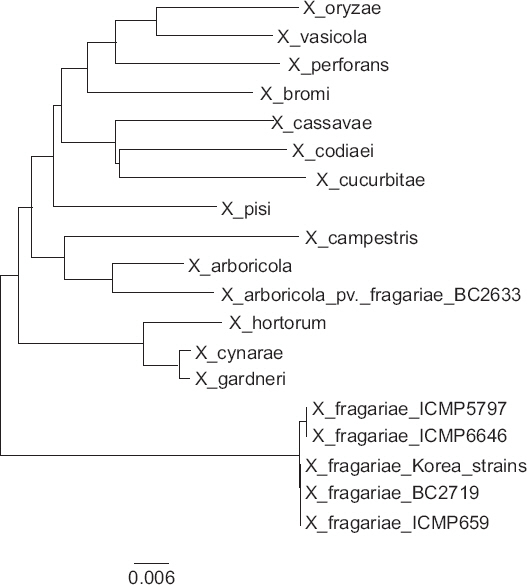
Fig. 2
Phylogenetic tree constructed by MEGA software on the basis of the concatenated sequence of the four housekeeping gene fragment dnaK, fyuA, gyrB, and rpoD.
국내에서 분리된 딸기세균모무늬병균들의 조사된 생리생화학반응은 차이가 없었다. X. fragariae의 종 내 변이를 생리생화학반응(Van den Mooter와 Swings, 1990), 지방산(Janse 등, 2001; Roberts 등, 1998), 단백질(Janse 등, 2001), restriction fragment length polymorphism (Roberts 등, 1998), repetitive-sequence PCR (Opgenorth 등, 1996; Stöger 등, 2008), random amplfied polymorphic DNA PCR (Pooler 등, 1996), MLSA (Young과 Park, 2007) 등으로 분석하였으나 종 내에 뚜렷한 차이를 보이지 않았다. 한국에서 분리된 X. fragariae의 조사된 생리생화학반응 특성은 Van den Mooter와 Swings (1990)의 결과와 X. fragariae의 대표 균주(LMG 708T)의 특성과 일치하였고, 딸기세균잎마름병균(X. arboricola pv. fragariae CFBP 6771PT)과 뚜렷한 차이를 보였다(Table 3). 분리된 세균들은 Gram 음성이었고, YDC 배양기에서 점액질 성장, 전분가수분해, 빙핵형성 등은 양성반응을 보였고, 35°C에서 성장, SX 배양기에서 성장, esculin 가수분해, 단백질 분해 arabinose를 이용한 산생성, 성장에 글리세롤과 melibiose 이용 등은 음성반응을 보였다. 그리고 litmus milk에서 알카리를 생성하였다(Table 3).
사용된 유전자에 따라 세균의 계통수분석으로 세균 스트레인들의 분화정도를 다르게 표현할 수 있다(Ait Tayeb 등, 2005). Young 등(2007)은 Xanthomonas속 계통분류에서 사용된 fyuA, dnaK, gyrB, rpoD 유전자들은 동일 종의 스트레인들을 구별할 수 있었다. 본 연구에서 fyuA, dnaK, gyrB, rpoD 유전자를 사용하여, 국내에서 분리된 딸기세균모무늬병균의 항존유전자 염기서열의 다양성을 분석하였다. 조사된 딸기세균모무늬병균의 dnaK (940 bp), fyuA (698 bp), gyrB (865 bp), rpoD (873 bp) 유전자들은 X. fragariae 대표 균주의 각 유전자의 염기서열과 동일하였다. 국내에서 분리된 X. fragariae의 gyrB, fyuA 유전자는 GenBank에 등록된 X. fragariae ICMP 5797과 ICMP 6646 세균과 100% 일치하였으나, dnaK, rpoD 유전자는 각 유전자에 대하여 1염기 차이를 보이며 99.9% 일치하였다. dnaK, fyuA, gyrB, rpoD 유전자들을 순서대로 배열한 3,374 bp 염기의 MLSA 염기서열을 사용한 계통수를 분석하였다. 비록 사용된 병원세균의 수가 제한적이지만 계통수 분석에서 Young 등(2007)의 결과와 같이 사용된 X. fragariae는 2종류의 스트레인으로 구별되었다. 국내 분리균은 딸기세균잎마름병균인 X. arboricola pv. fragariae와 다른 그룹에 포함되었고, X. fragariae 대표 균주와 동일 그룹에 포함되었다(Fig. 2).
딸기세균모무늬병 화학적 방제를 위하여 항생제(Alippi 등, 1989)와 동제화합물(Jones 등, 1991)을 사용하고 있으나 시간경과에 따라 방제효율이 제한적이고(Stall과 Thayer, 1962), 약해를 유발시킬 수 있어(Howard와 Albregts, 1973) 저항성 품종 재배가 추천된다. 외국에서 상업용 딸기재배 품종 중 딸기세균모무늬병 저항성 품종은 알려져 있지 않다(Smith 등, 1992). 딸기 유전자원 중 저항성을 보이는 F. virginiana (US4808)와 F. virginiana × F. × ananassa 교배형(US4809)이 보고되어 있으나(Maas 등, 2000), 딸기품종들은 딸기세균모무늬병에 감수성이 다양한 것으로 보고되었다(Desmet 등, 2009). 최근 연구에서 스페인에서 재배되고 있는 딸기품종 중 ‘Sieger’가 X. fragariae에 저항성으로 보고되었으나(Pérez-Jiménez 등, 2012) 현재까지 상업적으로 생산되는 딸기품종들은 딸기세균모무늬병에 감수성으로 알려져 있다(Bestfleisch 등, 2015). 국내에서 재배되고 있는 딸기품종들의 딸기세균모무늬병에 대한 저항성 조사 결과 국내에서 재배되고 있는 금향, 싼타, 숙향 등 저항성 평가에 사용된 18품종들은 감수성 품종으로 알려진 스위트 챨리(Sweet Charlie; Lewers 등, 2003)와 같이 감수성이었다(Table 4). 모든 품종들은 접종 3일부터 접종 위치가 수침상으로 변하고 습도가 높은 오전에 삼출물이 형성되었다. 1주일 후부터 병환부는 괴저증상으로 변하고 확대되었다. 이상의 결과는 Maas 등(2000)의 저항성 등급을 근거로 국내에서 재배되는 딸기품종들의 저항성 등급 4와 일치하였다.
Table 4
Strawberry variety reactions to infection by strains BC3191 and BC3195 of Xanthomonas fragariae in tests 1 and 2, and an overall rating two week after inoculation
| Strawberry variety tested | Disease reaction | Overall rating | |||
|---|---|---|---|---|---|
|
|
|||||
| Test 1 | Test 2 | ||||
|
|
|
||||
| BC3191 | BC3195 | BC3191 | BC3195 | ||
| Geumhyang | 4* | 4 | 4 | 4 | S |
| Daewang | 4 | 4 | 4 | 4 | S |
| Dochinimine | 4 | 4 | 4 | 4 | S |
| Dochiodome | 4 | 4 | 4 | 4 | S |
| Red Pearl | 4 | 4 | 4 | 4 | S |
| MaeHyang | 4 | 4 | 4 | 4 | S |
| Benihotbe | 4 | 4 | 4 | 4 | S |
| Bogyojosaeng | 4 | 4 | 4 | 4 | S |
| Sajjinoka | 4 | 4 | 4 | 4 | S |
| Seolhyang | 4 | 4 | 4 | 4 | S |
| Suhong | 4 | 4 | 4 | 4 | S |
| Sukhyang | 4 | 4 | 4 | 4 | S |
| Sweet Charlie | 4 | 4 | 4 | 4 | S |
| Ssanta | 4 | 4 | 4 | 4 | S |
| Akihime | 4 | 4 | 4 | 4 | S |
| Okmae | 4 | 4 | 4 | 4 | S |
| Jukhyang | 4 | 4 | 4 | 4 | S |
| Festival | 4 | 4 | 4 | 4 | S |
* Resistance rating based on Maas et al. (2000), two weeks after infiltration to leaves with the bacterial strains, BC3191 and BC3195.
 PDF Links
 PubReader
 Full text via DOI
 Download Citation
 Print
Print


