


Deltoid synurus (Synurus deltoides) belongs to the family Asteraceae and is distributed in China, Japan, Korea, and southern Siberia (Plants of the World Online, 2022). The plant grows in the mountains and is cultivated as a wild vegetable and medicinal plant in Korea. Foot rot symptoms were repeatedly observed on plants of deltoid synurus growing in a field of the Wild Vegetable Research Institute located in Pyeongchang, Korea during disease surveys in July 2020 and June 2021. The symptoms appeared as wilting of the plant leaves, and the plant stems and petioles at or above the soil line turned dark and rotted (Fig. 1AŌĆÆ D). Severely diseased plants wholly wilted and blighted. Two sites were observed in the field, and twenty plants at each site were investigated for the disease incidence during the disease surveys. The incidence of diseased plants in the field was 5ŌĆÆ10%.
Fig.┬Ā1.
Foot rot symptoms in deltoid synurus plants. (AŌĆÆ D) Symptoms observed in the field investigated. (E, F) Symptoms induced by artificial inoculation tests with Phytophthora cryptogea isolates. (G) A non-inoculated plant (control).

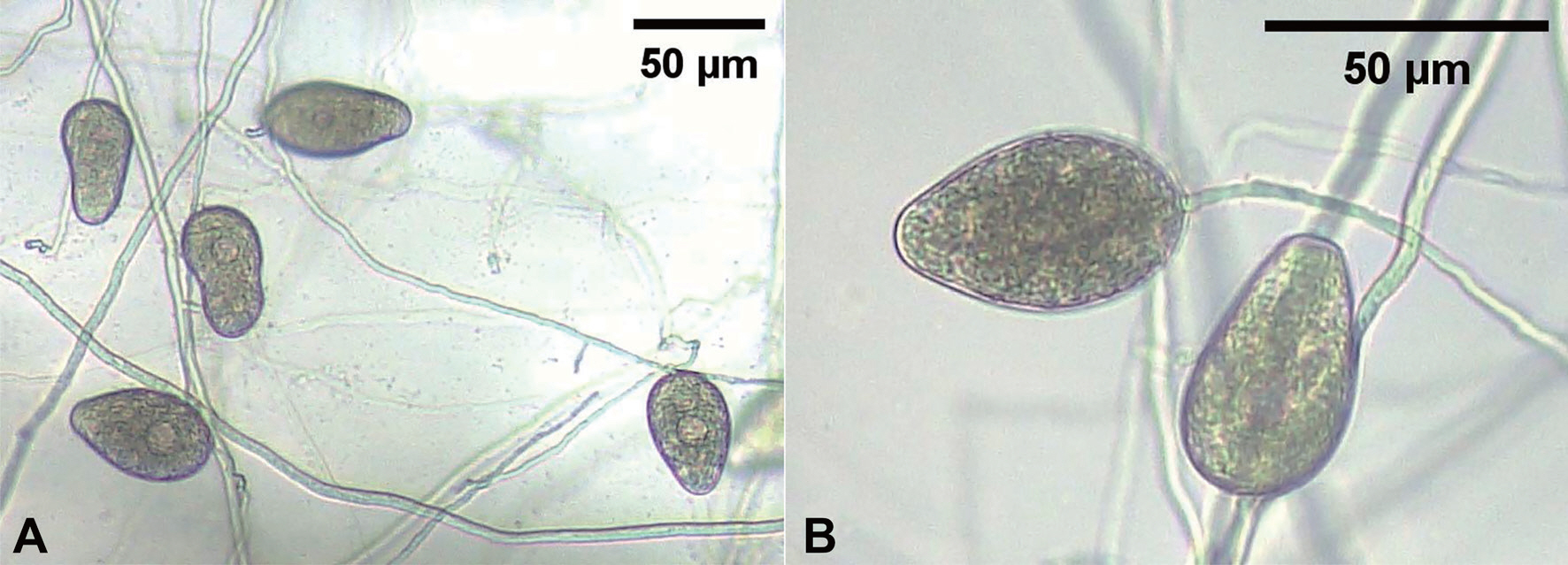
Fungal pathogens were isolated from diseased plants of deltoid synurus. The 3-5 mm long lesion pieces cut from stems of the diseased plants were plated on 2% water agar after surface-sterilizing with 1% sodium hypochlorite solution for one min. Single hyphal tips growing from the lesion pieces were transferred to potato dextrose agar (PDA) slants after incubating the plates at 25┬░C for 2 days. Five isolates of Phytophthora sp. were obtained from the lesion pieces. Each isolate was cultured on V8-juice agar (V8A) in 9 cm diameter Petri dishes at 25┬░C in the dark for 7 days. Eight mycelial disks of 10 mm in diameter were cut and removed from the V8A cultures. Ten milliliters of sterile distilled water was poured into the culture dishes, and the dishes were incubated at 25┬░C under fluorescent light for 7 days. Twenty to thirty sporangia per isolate produced inside the eight mycelial disk holes in the culture dishes were examined for their morphological characteristics using a compound light microscope (Nikon Eclipse Ci-L, Tokyo, Japan). Sporangia were oval to obpyriform with rounded bases, nonpapillate, persistent on the sporangiophores (Fig. 2A, B), and measured 37.8ŌĆÆ66.2├Ś22.7ŌĆÆ36.3 ╬╝m (av. 53.0├Ś30.9 ╬╝m). The morphological characteristics of the isolates were similar to those of Phytophthora cryptogea described in a previous study (Ho and Jong, 1991).
Fig.┬Ā2.
Morphological features of Phytophthora sp. isolates from diseased deltoid synurus plants. (A, B) Hyphae, sporangiophores, and sporangia of the isolates.
To validate the morphological identification of the five isolates, DNA sequencing of translation elongation factor 1 alpha (ELF1╬▒) and beta-tubulin (╬▓-tub) genes was analyzed. Mycelia were harvested from 5- to 14-day-old PDA cultures of each isolate, and total genomic DNA was extracted from the mycelia using the CTAB methods (Zhang et al., 2010). ELF1╬▒ and ╬▓-tub genes were amplified from the genomic DNA using primer sets of ELF1╬▒F/ELF1╬▒R and ╬▓-tubF/╬▓-tubR, respectively (Van't Klooster et al., 2000; Weerakoon et al., 1998). The thermocycle sequence was as follows: an initial denaturation at 95┬░ C for 3 min; 30 cycles consisting of denaturation 95┬░ C for 30 sec, annealing temperatures were 60┬░ C for ELF1╬▒ and ╬▓-tub. The PCR products were cloned into T-blent vector (Solgent Inc., Seoul, Korea) and sequenced (Macrogen Inc., Seoul, Korea). The obtained sequence data were compared to Phytophthora spp. strain sequences available in the NCBI GenBank database. The phylogenetic tree construction was carried out using MEGA11 in the bootstrap of 1,000 by the maximum likelihood method and Kimura 2-parameter model (Tamura et al., 2021). The tree was constructed based on concatenated sequences of two gene fragments in ELF1╬▒ and ╬▓-tub re-gions of Phytophthora spp. The phylogenetic tree based on loci ELF1╬▒ and ╬▓-tub combined sequence data showed that all the isolates were clustered in a group with P. cryptogea strains (Fig. 3). The nucleotide sequences of ELF1╬▒ and ╬▓-tub genes obtained from the five isolates were deposited in NCBI GenBank with accession numbers of OM654412ŌĆÆ OM654416 and OM654417ŌĆÆ OM654421, respectively.
Fig.┬Ā3.
Maximum likelihood phylogenetic tree based on a concatenated alignment of the translation elongation factor 1 alpha and beta-tubulin sequences of five Phytophthora cryptogea isolates (SDPHY-2001ŌĆÆ SDPHY-2005) from deltoid synurus and reference species. The reference sequence data were obtained from the NCBI GenBank database. Statistical support for the branches was as-sessed by bootstrap with 1,000 replicates. Bootstrap values Ōēź50 are shown near the branch nodes.

In this study, five isolates of Phytophthora sp. from the diseased plants of deltoid synurus were identified as P. cryptogea based on the morphological and molecular characteristics. Among the five isolates of P. cryptogea, three were tested for pathogenicity on deltoid synurus plants using artificial inoculation. Each isolate was cultured on V8A to prepare the inoculum. Twenty mycelial disks (10 mm in diameter) cut from 7-day-old V8A cultures were transferred into 9 cm diameter Petri dishes with 20 ml of sterile distilled water and incubated at 25┬░C under fluorescent light for 7 days to produce sporangia. The Petri dishes were placed in the refrigerator at 4┬░C for 2 hr to induce release of zoospores from sporangia. Thereafter, the culture solution in the Petri dishes was filtered through four layers of gauze to prepare zoospore suspension. The concentration of each zoospore suspension was adjusted to 105ŌĆÆ106 zoospores/ml. The zoospore suspension of each isolate was inoculated to 75-day-old deltoid synurus plants that were grown in circular plastic pots (height, 15 cm; upper diameter, 17 cm; lower diameter, 10 cm) in a vinyl greenhouse. For inoculation test, surface soil around the plants in the pots was dug at a depth of 1ŌĆÆ2 cm. The 60 ml of each zoospore suspension was poured around the plant in a pot, and the inoculated plant part was covered with the original soil. The same quantity of sterile distilled water was used for the control plant. The inoculated plants were cultivated in a greenhouse at 24-30┬░ C. The inoculation test was performed in triplicate. Pathogenicity of the isolates was rated based on the degree of foot rot symptoms induced 10 days after inoculation.
The tested isolates of P. cryptogea induced foot rot symptoms in the inoculated plants (Fig. 1E, F), whereas no symptoms were observed in the control plant (Fig. 1G). The symptoms induced by the artificial inoculation of plants were similar to those observed in the diseased plants in the field investigated. The isolates that induced symptoms on the plants were re-isolated from the lesions.
P. cryptogea was considered to be synonymous with Phytophthora drechsleri (Ho and Jong, 1986). However, it has been reported that the two species to be distinct based on molecular analysis (Cooke et al., 2000). P. cryptogea has been reported to cause damping-off, foot rot, stem rot, leaf rot, and wilt in plants of 141 genera in 49 families (Farr and Rossman, 2022). However, there has been no report on disease occurrence caused by the pathogen in deltoid synurus. This is the first report of P. cryptogea causing Phytophthora foot rot in deltoid synurus.
 PDF Links
 PubReader
 ePub Link
 Full text via DOI
 Download Citation
 Print
Print


