First report of Cucumber mosaic virus in African Impatiens (Impatiens walleriana) in Korea
Article information
Abstract
Virus-like symptoms including stunt, severe mosaic with malformation of leaves, fern-like leaves and abnormal petals were observed from an African impatiens (Impatiens walleriana) grown in a plant nursery in Icheon, Korea. Serological analysis using immuno-strip kits for viruses reported in African impatiens indicated that Cucumber mosaic virus (named CMV-Im) was a causal agent for the symptomatic African impatiens. Biological properties of CMV-Im were analyzed using responses of host plant species, suggesting that CMV-Im is a typical strain that belongs to CMV subgroup I. RT-PCR analysis verified CMV-Im infection from naturally infected African impatiens or mechanically inoculated some host species. Analysis of multiple alignments of CMV capsid protein (CP) sequences showed that CMV-Im shared high CP amino acids identities with other CMV strains. Phylogenetic tree analysis for the CP sequences of CMV-Im and representative CMV strains confirmed that CMV is a typical member of CMV subgroup I. To our knowledge, it is the first report of CMV in African impatiens in Korea.
Impatiens walleriana, commonly called ‘African impatiens’ or ‘New Guinea impatiens’, is a year-round outdoor blooming perennial, native to eastern Africa including Kenya, Tanzania and Mozambique. African impatiens can be bred easily by cuttings and the shoots also have high regenerating capacity after harvesting. African impatiens is frequently used for bedding and decoration of borders or used as pot plants in the world. Since 1990’s, African impatiens plants have been grown in some gardens of Korea and have been distributed throughout the nation by commercial plant nurseries.
Cucumber mosaic virus (CMV), a type species of the genus Cucumovirus in the family Bromoviridae, is a positive-sense single-stranded RNA virus with a tripartite genome (Palukaitis et al., 1992; Palukaitis and Garcia-Arenal, 2003). CMV strains can be divided into three subgroups, IA, IB and II (Roossink et al., 1999; Palukaitis and Garcia-Arenal, 2003). CMV is consisted of three genomic RNAs. RNA 1 encodes 1a protein that contains domains of methyltransferase and helicase for viral replication. RNA2 encodes 2a protein (the viral polymerase) and 2b protein that is essential for viral systemic movement and infectivity in some host plants (Ding et al., 1995; Hayes and Buck, 1990). RNA 3 encodes the 3a protein (movement protein, MP) for viral movement and the capsid protein (CP) for encapsidation which is translated from a subgenomic RNA 4. CMV has an extremely broad host range of approximately 1,000 species including vegetables, cereals, flowering plants and trees (Gal-On et al., 1994; Lee et al., 2007; Palukaitis and Garcia-Arenal, 2003; Ryu et al., 1998).
In March 2014, virus-like symptoms including stunt, severe mosaic with malformation of leaves, fern-like leaves and abnormal petals were observed from an African impatiens plant grown in a plant nursery in Icheon, Korea (Fig. 1). The symptomatic leaves of African impatiens were analyzed for the presence of several ornamental viruses including Cucumber mosaic virus (CMV), Pepper mild mottle virus (PMMoV), Pepper mottle virus (PepMoV), Tobacco mosaic virus (TMV), Tomato bush stunt virus(TBSV), and Tomato spotted wilt virus (TSWV) by immuno-strip diagnostic kits that were developed in our laboratory. Positive controls and an extract from healthy leaves of African impatiens as a negative control were included in each immuno-strip assay. CMV was detected serologically from the naturally infected African impatiens but PMMoV, PepMoV, TMV, TBSV and TSWV were not detected from the African impatiens (data not shown). To further exclude the possibility of infection with Impatiens necrotic spot virus (INSV), the same positive controls and extract from healthy leaves of African impatiens were analyzed using a commercial immuno-strip for INSV (Agdia, USA), showing negative for INSV. These serological results suggest that CMV was a causal virus for the symptomatic African impatiens.
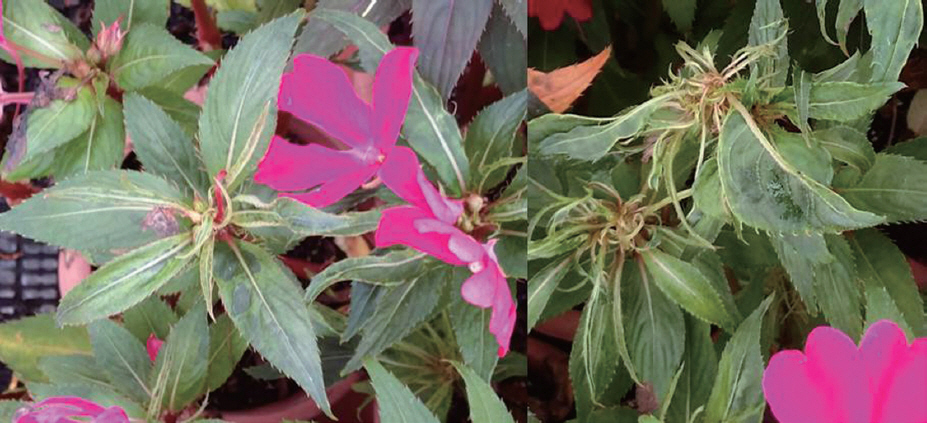
Foliar symptoms of the naturally infected African impatiens. Virus-like symptoms was observed in an African impatiens plant that grown in a plant nursery in Icheon, Korea.
To verify further whether CMV was a major agent for the disease symptoms, sap extract of the symptomatic African impatiens was mechanically inoculated to indicator plant species, Chenopodium quinoa, Datura stramonium, Nicotiana benthamiana, N. tabacum (cv. Xanthi-nc), and African impatiens (I. walleriana). Local lesions on the inoculated leaves of C. quinoa were produced and systemic mosaic symptoms was observed in upper leaves of N. benthamiana at 5 to 7 days postinoculation (dpi) (Fig. 2A and data not shown). Systemic mosaic symptoms on upper leaves of D. stramonium and N. tabacum (cv. Xanthi-nc) were observed at 10 dpi (Fig. 2B and 2C), respectively. African impatiens produced systemic mosaic and distortion on leaves from 30 dpi (Fig. 2D) and color-breaking symptoms on flowers at 60 dpi (Fig. 2E). Subsequently, CMV infections in all the symptomatic leaves of the inoculated indicator plants were verified using the CMV-immuno-strip kit (Fig. 3A).

Symptoms on leaves and flowers of the indicator plant species infected with CMV-Im. (A) Necrotic local lesions on the inoculated C. quinoa leaves at 5 dpi, (B) Systemic mosaic and fern-like symptoms on upper leaves of D. stramonium at 10 dpi, (C) Systemic mosaic symptom on upper leaves of N. tabacum (cv. Xanthi-nc) at 10 dpi, (D) Systemic severe mosaic on upper leaves of African impatiens inoculated mechanically with CMV-Im, (E) Color-breaking symptoms on petals of African impatiens inoculated with CMV-Im at 60 dpi.

Serological diagnostic assays and RT-PCR analysis for CMV detection. (A) Immuno-strip assay for CMV diagnosis from naturally infected African impatiens or mechanically inoculated with CMV-Im. Sap extracts from systemic leaves of D. stramonium, N. benthamiana, and N. tabacum (cv. Xanthi-nc) inoculated with CMV-Im were analyzed using immune-strip kits for CMV. A sap extract from leaf tissues of healthy African impatiens were used as a negative control. Sample names used for serological assays were indicated on the top. Control line and test line for CMV-immuno strip kits were indicated on the right of the panel. (B) A representative RT-PCR analysis for CMV detection. CMV-Im was detected from the naturally infected African impatiens and CMV-Fny was detected from an upper leaf of the infected tobacco plant. Total RNAs were extracted using a commercial RNA purification kit (Qiagen, Germany). Total RNA from leaf tissues of healthy African impatiens were used as a negative control. The complete full-length cDNA of CMV CP gene was indicated on the right as an arrow. M, 1000 bp DNA size marker indicated on the left.
To determine whether the strain of CMV from African impatiens belongs to CMV subgroup I, based on strong positive reactions in CMV-immuno strip kits, reverse transcription (RT)- PCR was performed using CMV-specific primers. CMV Fny strain was used as a positive control (Palukaitis et al., 1992) for comparison. Total RNAs were extracted using the RNeasy Plant Mini Kit (Qiagen, Germany), according to the manufacturer’s instructions. RT-PCR was carried out using the One-Step® RT-PCR kit (Invitrogen, USA) using specific primers (CMVCPFor, 5´-ATGGACAAATCTGAATCAACC-3´ and CMVCPRev, 5´-TTCAGACTGGGAGCACTCCAG-3´), which was designed to amplify the complete full-length CMV CP gene. The thermocycling conditions were as follows: 60 min at 50°C for RT, 5 min at 95°C (1 cycle), 30 sec at 94°C, 30 sec at 55°C, and 1 min at 72°C (35 cycles), and a final extension for 10 min at 72°C. RT-PCR products were analyzed in 1.2% agarose gel and visualized after soaking in ethidium bromide (EtBr) solution. As expected, RTPCR products of approximately 660 bp were synthesized from the naturally infected African impatiens and a tobacco plant infected with CMV Fny strain (Fig. 3B), meanwhile none of RT-PCR product was amplified from a healthy African impatiens. These results confirm that a strain of CMV (named CMV-Im) was an authentic causal virus in the severe disease of African impatiens. The amplified RT-PCR product of CMV-Im was directly cloned into the pGEM-T Easy vector (Promega, USA) to determine nucleotide sequence of the complete CP gene, according to the manufacturer’s instruction. Subsequently, transformation to E. coli and selection of cDNA clones using EcoRI digestion were performed according to standard protocols. Three individual cDNA clones were analyzed for sequencing using the BigDye terminator cycle sequencing kit, according to the manufacturer’s instructions (Applied Biosystems, USA). The complete nucleotide sequence of the CMV-Im CP gene is 657 bp long (GenBank accession No. LC066587) and the molecular mass of CMV-Im CP from deduced amino acids is 24.12 kDa consisting of 218 amino acids. The CP nucleotide and amino acid sequences of CMVIm shared 76.0 to 93.3% nucleotide identity and 82.1 to 98.6% amino acid identity with thoses of other CMV isolates (Table 1).
Sequence identities of nucleotide (nt) and the deduced amino acid (aa) between CMV-Im and other representative CMV strains
Pairwise alignments with Bootstrap analyses of 1,000 replicates were performed to evaluate the significance of the interior branches. The phylogenetic tree of CMV isolates produced using the alignment of the CP amino acid sequences is presented in Fig. 3. The phylogenetic tree constructed from CP sequences of 12 CMV strains revealed three clades (IA, IB and II) of the isolates into two monophyletic clusters as described previously (Roossink et al., 1999; Palukaitis and Garcia-Arenal, 2003). Interestingly, the phylogenetic tree analysis of the CPs suggests that the CMV-Im belongs to subgroup IB and was closer the CMV-Imp isolated from Impatiens in China than other CMV Impatiens isolates (Fig. 4). It remains to be determined if CMV-Im is a member of subgroup IB based on sequence comparisons of 5´-nontranslasted region of RNA3 (Roosinck et al., 1999).

Phylogenetic tree analysis for CPs of CMV-Im and representative CMV strains, based on the deduced CP amino acid sequences. Multiple sequence alignments were analyzed using DNAstar software and phylogenetic tree was constructed from calculations from pairwise amino acid sequence distances derived from multiple-alignment format. The accession numbers of these CMV strains were listed in Table 1. Taxonomical classification of each CMV strain reported previously was indicated on the right.
Our results suggest that members of CMV subgroup IA and IB are some of the important viruses to threat for production of African impatiens in Korea and other countries. We showed that CMV-Im caused severe mosaic symptoms of leaves and colorbreaking symptoms on flowers of African impatiens, indicating CMV infection is likely to cause decline in quality of flowers for many ornamental crops including African impatiens. African impatiens infected with CMV in plant nurseries during season of seedling production can be a source of CMV for epidemic virusdisease of African impatiens by transmission of aphid vectors. Therefore, effective controls of aphid vectors are a good strategy for CMV management in African impatiens as well as other ornamental crops since CMV is a non-persistently transmitted virus. To our knowledge, this is the first report of CMV in African impatiens in Korea.
Acknowledgement
This study was supported by a grant of Basic Research Program (PJ008472022015) of National Institute of Horticultural and Herbal Science, RDA, Republic of Korea.