Res. Plant Dis > Volume 21(3); 2015 > Article
Chu, Shin, Park, and Jeong: Effect of Gamma Irradiation on Botrytis cinerea Causing Gray Mold and Cut Chrysanthemum Flowers

ABSTRACT

Gray mold caused by Botrytis cinerea is one of the most important postharvest fungal pathogens of cut flowers. Here, gamma irradiation, an alternative for phytosanitary purposes, and sodium dichloroisocyanurate (NaDCC) were used to control B. cinerea in a cut chrysanthemum (Chrysanthemum morifolium Ramat.) cultivar, ‘Baekma’, one of the cultivars susceptible to B. cinerea. Spore germination and mycelium growth of B. cinerea were inhibited by gamma irradiation in an inversely dose-dependent manner. A dose of 4 kGy completely inhibited the mycelium growth of B. cinerea. A significant change in flower quality (physical properties) on chrysanthemum was shown from gamma irradiation at over 0.2 kGy (p<0.05). Therefore, in this study, the integration of gamma ray (below 0.2 kGy) and NaDCC, an eco-friendly form of chlorine, was investigated to control the disease with low dose of gamma irradiation dose. Interestingly, the gamma irradiated flowers showed more disease severity than the non-irradiated flowers. The combined treatment of gamma irradiation and NaDCC does not affect the severity of the fungal disease, whereas only 70 ppm of NaDCC treatment showed a significantly reduced severity. These results suggest that only chlorination treatment can be applied to control B. cinerea in cut chrysanthemum flowers.

Introduction

Chrysanthemum (Chrysanthemum morifolium Ramat.) is one of the most popular floricultural (cut-flower) and ornamental (pot and garden) crops in the world, along with roses, carnation, and more recently, gerbers. Cut chrysanthemum has a great ornamental value, making up 30% of all cut flowers globally (Silva, 2003). Botrytis rot, caused by the serious fungal pathogen Botrytis cinerea Pers., is a widespread disease of ornamental crops, flowers, and vegetables (Schwinn, 1992). The disease is favored by an extended period of cloudy, humid (relative humidity; RH above 93%) conditions, and wet weather (Williamson et al., 1995). B. cinerea infections often were not able to detect at the time of harvest, but develop rapidly under moist conditions during storage and shipment (Vrind, 2005). Once infected, petals during wet or humid weather, show masses of silver-gray spores on the whole petals, resulting in a collapse of the flower head and petal drop (Elad, 1998). Currently, chemical fungicides are being used either as pre-harvest sprays in a greenhouse or as postharvest dips of cut flowers to prevent B. cinerea. However, latent infections are not efficiently controlled by fungicide treatments (Elad, 1998). In addition, the continued use of chemical fungicides causes fungal resistance to fungicides and chemical residues (Williamson et al., 2007). CO2 atmosphere, photochemical, and calcium sulfate have been used to control B. cinerea on cut chrysanthemum flowers as alternative treatments (De Capdeville et al., 2005; Hammer et al., 1990). However, these methods only in completely inactivate fungal pathogens. Therefore, it is necessary to develop a new method to increase the vase-life of cut chrysanthemum as well as to control postharvest disease.
The use of irradiation accelerated five decade ago with safety, efficacy, and feasibility. Irradiation can effectively control microorganisms and insects that can cause spoilage or damage to fresh commodities. It is a fitting method for controlling the pathogens on fruits, fresh-cut vegetables, flowers, and seeds. It is not only used on the surface, but can also penetrate the product and inactivate pathogens that exist in products. Following increasing concerns about the use of fungicides, irradiation is being reconsidered as a viable alternative and effective nonchemical treatment for control of postharvest diseases (Hallman, 2011). So far, most extensive research with irradiation has been performed on insects to prevent the development of reproduction on imported or exported horticultural products from low-dose irradiation. Although doses of as low as 0.4 kGy are sufficient to sterilize most harmful insects, it is impossible to fully control postharvest fungal diseases of cut flowers (Blank and Corrigan, 1995). The radiation doses required to completely inactivate fungi alter certain physiological properties, such as leaf drop and bent neck of cut flowers (Fjeld et al., 1994). Thus, irradiation can be used in combination with other treatments, such as heat, natural compounds, physical treatments, and eco-friendly agents, to reduce the irradiation dose, thereby increasing the effectiveness, while reducing the negative effect (Bustos-Griffin et al., 2012). One of the promising treatments with irradiation to disinfect postharvest diseases is chlorination. Chlorination damages microbe cell membranes, proteins and nucleic acid through oxidative degradation (Dychdala, 1983). This is effective, relatively inexpensive, and non-residual chemical, which is widely used to reduce bacterial and fungal diseases on fruit and vegetable surfaces and processing equipment (Suslow, 1997). A recent study showed that combined treatment of gamma irradiation with dichloroisocyanurate (NaDCC) effectively inhibited the development of a fungal pathogen (Yoon et al., 2014).
In the present study, individual and combined treatments were conducted to evaluate inhibition of B. cinerea on cut chrysanthemum flowers. The effect of gamma irradiation on the physiological responses of fungal spores and cut chrysanthemum flowers was evaluated. Moreover, we also determined the optimal condition for the control of postharvest B. cinerea of cut chrysanthemum flowers. The aim of the present study was to develop a practical solution for the control of the B. cinerea infection of cut chrysanthemum flowers.

Material and Methods

Source of material and production of inoculum

Botrytis cinerea was obtained from Korean Agricultural Culture Collection (KACC40573). For a pure culture, isolated individual conidia were transferred to potato dextrose agar (PDA, Difco Laboratories, Detroit, MI). Prior to each experiment, all cultures were grown on PDA at 25°C for 14 days. Cultures were transferred to a fresh PDA every 14 days. To harvest the conidia, about 10 ml of sterile distilled water (SDW) was added to a culture Petri dish and the conidia were gently harvested by filtration through four layers of gauze. The conidial suspensions were collected in sterile screw-cap test tubes (50 ml) containing 15 ml of SDW and filtered twice using sterile Pasteur pipettes packed with glass wool (Saleh et al., 1988). The concentration of conidia was measured using a hemacytometer (Warner-Lambert Technologies Inc., Buffalo, N.Y.). The final conidial concentration was adjusted to 105 conidia ml-1 with SDW for the experiments.

Gamma irradiation

A cobalt-60 gamma irradiator at the Korea Atomic Energy Research Institute, Jeongeup, Korea (150 TBq capacity; ACEL, MDS Nordion, Canada) was used for the irradiation. All of the absorbed doses were calibrated using alanine dosimeters with a diameter of 5 mm (Bruker Instruments, Rheinstetten, Germany), where a Bruker EMS 104 EPR analyzer (Bruker Instruments, Rheinstetten, Germany) was used to determine the free-radical signals.

NaDCC treatment

For chlorination, the cut chrysanthemum were sprayed with sodium dichloroisocyanurate (NaDCC; Sigma-Aldrich, Poole, UK) at various concentrations (From 30 to 70 ppm) 1 h prior to irradiation.

Experiments with pure cultures

The viability of irradiated-spores was determined by serial dilution plate method, then incubated at 25°C for 3 days (Acquino et al., 2005). Survival curves were created by exponential function to the survivor CFU/ml versus the actual radiation dose. For inhibition of fungal mycelium, agar discs (5 mm in diameter) were isolated from a solid culture of irradiated B. cinerea, and each agar disc was placed on a PDA medium (90 mm in diameter). After incubation at 25°C in dark for 3 days, mycelial growth of B. cinerea was measured as an average colony radius.
For an assay of spore germination, spores (105 conidia ml-1) were treated with 0, 0.5, 1, 2, 3, and 4 kGy of gamma irradiation and then incubated for the times indicated at 25°C. Under a microscope (40×), for every zone selected, germinated and non-germinated spores were counted (Eclipse Ni, Nikon, Japan). Ten representative zones were selected at random. The sum of germinated spores over the total spores present determined the percentage of germination for B. cinerea at the time of sampling.
For scanning electron microscope (SEM) analysis, irradiated fungal spores were fixed in 2% OsO4 for 1 h, dehydrated in acetone. The dehydrated spores were dried by incubating in hexamethyldisilazane (HMDS) for 15 min twice, mounted on carbon tape, coated with platinum, and then examined under a SEM (JEOL, Tokyo, Japan).
Zero point of 5 milligram of freeze-dried mycelium of gamma-irradiated B. cinerea was added to 50 ml of a phosphate buffer with pH 8.0, then left over night at room temperature and filtered, and the filtrate was used for measuring proteins and total soluble sugars. The quantification of released proteins and total soluble sugars were determined by the Bradford assay and the phenol-sulfuric method (Bradford, 1976; Dubosis et al., 1956). The experiment was repeated twice.

Measurement of cellular leakage

The leakage of cytoplasmic contents from the mycelia of B. cinerea irradiated with gamma irradiation (600 Gy/h) was measured according to previous study with some modifications (Lewis and Papavizas, 1987). B. cinerea was cultured in a PDB (Potato Dextrose Broth) medium on a rotary shaker at 100 rpm at 25°C for 3 days. The mycelia were then harvested and washed with SDW. The washed mycelia were irradiated with 0, 0.5, 1, 2, 3, and 4 kGy, and incubated on a rotary shaker for 1, 2, 3, and 4 h. The mycelia were filtered, and the filtered solutions were used for determining the leakage of soluble proteins and soluble sugars. The quantification of released proteins and total soluble sugars were determined by the Bradford assay and the phenol-sulfuric method (Dubosis et al., 1956). The experiment was repeated twice.

Experiments with flowers

A cultivar of cut chrysanthemum (C. morifolium Ramat.), ‘Baekma’, was obtained from a commercial grower. The flowers were harvested at the normal growth stage, when sepals begin to split and petals become visible. The stems were recut 30 cm below the receptacles, and all except two or three distal leaves were removed. Each experimental unit consisted of three flowers in a 500-ml bottle containing 200 ml of water.
After the cut chrysanthemum were irradiated with 0, 0.2, 0.4, 0.6, 0.8, and 1 kGy of gamma irradiation, the physical changes were monitored from phytotoxicity, water uptake, fresh weight, and flower opening. Phytotoxicity was observed at 7 days after gamma irradiation. For water uptake, the difference between consecutive weighings of the centrifuge tube plus solution (without the flower) was used to calculate the water uptake. The percentage change in water uptake relative to the initial weight was then calculated for each time point. Flower opening and fresh weight were recorded daily according to the method of Rattanawisalanon (2003). For the flower opening, the diameter of irradiated-cut chrysanthemum was measured at doses of 0.2, 0.4, 0.6, 0.8, and 1.0 kGy of gamma irradiation at 7 days. The vase life of an inflorescence was terminated when 50% of the open flowers had wilted or abscised. The fresh weight of whole flower stems was also determined after gamma irradiation at 7 days. The percentage of change in fresh weight relative to the initial weight was then calculated for each time point. The experiments were repeated three times with similar results.
Cut chrysanthemum flowers were inoculated by spraying with conidia (105 spores/ml) of B. cinerea to the point of incipient runoff. Non-inoculated controls, sprayed with distilled water, were included as negative control for disease levels. After inoculation of B. cinerea, the flowers were treated with NaDCC, gamma irradiation, and combined treatment. Then, the flowers were placed in humidified storage chambers (relative humidity ranged from 95 to 100%) at 25°C. The flowers were removed from storage 7-10 days after inoculation, and the disease severity (0-4, 0: no infection; 1: 1 to 10% leaf area infected; 2: 11 to 25% leaf area infected; 3: 25 to 50% leaf area infected; 4: 50 to 100% leaf area infected) was evaluated as the number of lesions on each flower.

Statistical analysis

Data were subjected to analyses of variance (ANOVA) at p<0.05 using SPSS 13.0 software for Windows (SPSS Inc., USA) and Duncan’s multiple range tests (DMRT) were used to compare the differences among the mean values.

Results

Effect of gamma irradiation on fungal viability

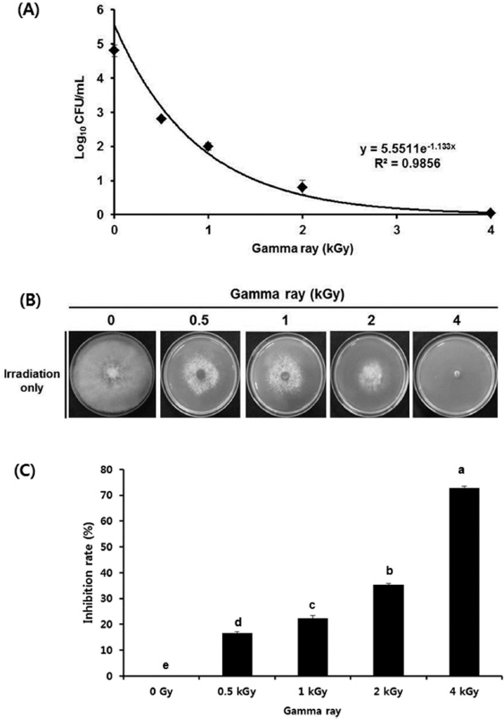
The viability of B. cinerea after gamma irradiation revealed that gamma irradiation significantly reduced the viability of fungal spores (Fig. 1A). Spores of B. cinerea decreased from 4.82±0.03 log10 CFU/ml (initial) to 0.89±0.06 log10 CFU/ml (2.0 kGy), whereas B. cinerea was not detected after 4.0 kGy of a gamma irradiation. The sensitivity of B. cinerea was significantly increased by dose-dependent gamma irradiation (p<0.05). Inhibition of the growth of fungal growth of gamma irradiation B. cinerea was evaluated through an inhibition assay of fungal mycelial growth on PDA medium with gamma irradiation. The degree of inhibition was noted as a percentage to the control, with 100% inhibition indicating complete inhibition. Gamma irradiation inhibited 0, 15, 22, 34, and 100% of the growth of B. cinerea at dose of 0, 0.5, 1.0, 2.0, and 4.0 kGy, respectively (p<0.05). Consistent with fungal spore viability to gamma irradiation, the mycelial growth of B. cinerea was completely inhibited at over a dose of 4.0 kGy. Inhibition of mycelial growth gradually increased at higher radiation doses, indicating that the inhibition of fungal development to gamma irradiation showed a dose-dependent manner (Fig. 1B, 1C).
Fig. 1
Sensitivity of gamma irradiation of B. cinerea of cut chrysanthemum flowers. (A) Survival curves of B. cinerea of cut chrysanthemum flowers. The applied doses in this study were 0, 0.5, 1.0, 2.0, and 4.0 kGy. Counts were recorded as log colony-forming units (log CFU)/ml sample. Experiments were conducted independently three times. The error bar is the standard errors of three replications. (B) Growth of B. cinerea after exposing with various doses of gamma irradiation (0, 0.5, 1.0, 2.0, and 4.0 kGy). (C) Changes in growth of B. cinerea measured at irradiation doses. Inhibition (%) means the percentage of fungal inhibition in comparison with untreated control of 0 kGy. The values followed by the different letters are significantly different (p<0.05) by Duncan’s multiple range test (n=3).
RPD_21_193_fig_1.jpg

Morphological alteration of B. cinerea spores to gamma irradiation

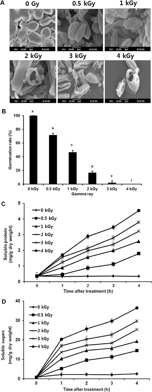
After confirming the inhibitory effects of gamma irradiation, morphological changes of fungal spores after irradiation were examined by analyzing the SEM images. SEM was employed with spores irradiated at a dose of 0.5, 1.0, 2.0, 3.0 and 4.0 kGy. It was obvious from the images that the shapes of higher dose irradiated-spores showed significant structural changes compared to non-irradiated spores (Fig. 2A). When spores were irradiated below 0.5 kGy, structural changes were not much different. However, a dose of over 1.0 kGy showed that the spores were crushed and exhibited a high degree of hollowness on the spore surface, whereas the non-irradiated spores were short, planiform elliptical rods. The present study demonstrated remarkable dose-dependent alterations in the surface of the B. cinerea spores. To expand the observation of the influence of irradiated fungal spores on the growth of B. cinerea, changes in spore germination were evaluated at 12 h incubation after irradiation. After gamma irradiation, 68.5%, 43.7%, 16.3%, 4.3%, and 0% of the spores were germinated after 12 h incubation at a dose of 0.5, 1.0, 2.0, 3.0, and 4.0 kGy, respectively while 98.9% was observed for the non-irradiated control (Fig. 2B). A significant decrease (p<0.05) in germination percentage of B. cinerea spores was found in a dose of 1.0-2.0 kGy. The significant inhibition of fungal growth due to irradiation was related to the physiological malfunction of spores caused by a loss of total protein (Geweely and Nawar, 2006). To verify the hypothesis regarding whether irradiation causes the cell death of B. cinerea, the leakage of the cytoplasmic contents was determined. As expected, irradiation significantly induced the leakage of soluble proteins and soluble sugars out of the mycelia of B. cinerea (Fig. 2C, D). These results confirmed previous findings that fungal spores were cracked and ruptured when exposed to increasing doses, indicating that irradiation possibly precipitated the plasma membrane disintegration and leakage of intracellular proteins and sugars, thus inhibiting the fungal growth.
Fig. 2
Effect of gamma irradiation on the spore structure and germination. (A) The surface morphology of fungal spores analyzed by SEM after gamma irradiation (0, 0.5, 1.0, 2.0, 3.0, and 4.0 kGy). (B) Microscopy image of B. cinerea spores after gamma irradiation. The images were taken after 12 h of incubation. Scale bar=20 μM (C) Relative spore germination rates observed after 12 h incubation after gamma irradiation. Values followed by the different letters are significantly different (p<0.05) by Duncan’s multiple test (n=3). (D) Total proteins were measured after gamma irradiation (p<0.05). The error bar is the standard errors of three replications.
RPD_21_193_fig_2.jpg

Effect of gamma irradiation on cut chrysanthemum quality

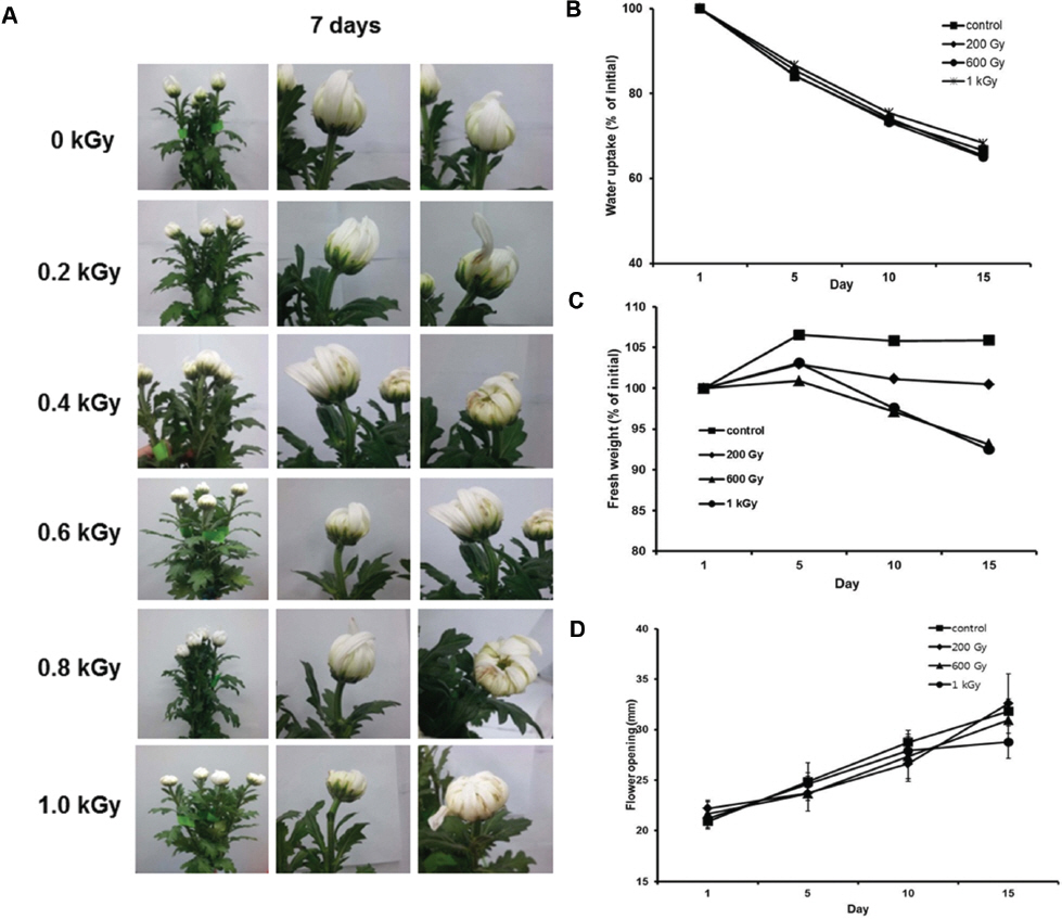
To obtain the minimal dose that does not affect the cut chrysanthemum quality to gamma irradiation, changes in physical properties of irradiated cut chrysanthemum was evaluated at various doses of gamma rays on a cultivar of cut chrysanthemum, ‘Baekma’. Cut chrysanthemum irradiated at over a dose of 0.2 kGy showed bleached or blackened areas at 7 days, 1-3 mm wide flowers, and a bent neck in ‘Baekma’ (Fig. 3A). The blackened areas and bent neck were not immediately noticeable after irradiation, but began to appear several days thereafter, but there were no other visible phytotoxicity symptoms on the flowers or foliage. This radiation-induced damage was more severe with increasing radiation dose and time in storage. As a result of the data obtained from the injury threshold experiment, irradiation dosages were decreased under 0.2 kGy, which was less injurious to the cut flowers for further study.
Fig. 3
Physical properties of irradiated cut ‘Baekma’ chrysanthemum. (A) The images showing the phytotoxicity of cut chrysanthemum after 7 days after various doses of gamma irradiation (0-1.0 kGy). (B) Changes in water uptake of cut ‘Baekma’ for 15 days after irradiation. The points are the means of four replicates±SD. (C) Fresh weight changes of cut chrysanthemum flowers after gamma irradiation and storage for 15 days at 10°C. (D) Changes in flower opening of ‘Baekma’ for 10 days after irradiation. The error bar was the standard error of three replications.
RPD_21_193_fig_3.jpg
The water uptake rates of ‘Baekma’ in water showed similar rates at doses of 0.2, 0.6, and 1.0 kGy of gamma irradiation for 15 days (Fig. 3B). The fresh weigh of chrysanthemum irradiated at 0.2, 0.6, and 1.0 kGy in water increased up to 5% at 5 days. Control and 0.2 kGy irradiated chrysanthemum maintained a similar fresh weight for 15 days. However, 0.6 kGy and 1.0 kGy irradiated chrysanthemum were dramatically decreased after 5 days (Fig. 3C). As in the fresh weight experiment, over the 0.2 kGy dosage, the chrysanthemum began to lose fresh weight faster, which shortened the vase life. For an evaluation of flower opening to gamma irradiation, ‘Baekma’ were irradiated at doses of 0.2, 0.6, and 1.0 kGy, and the length of the opened flowers was then measured. Flower opening of irradiated cut flowers was not inhibited in a dose-dependent manner (Fig. 3D). These physical responses to gamma irradiation result suggesting that a dose of 0.2 kGy is a threshold of irradiation on ‘Baekma’. It was interesting note that treatment of below 0.2 kGy also elevates the vase life of cut chrysanthemum.

Effect of gamma irradiation and NaDCC on B. cinerea in cut chrysanthemum

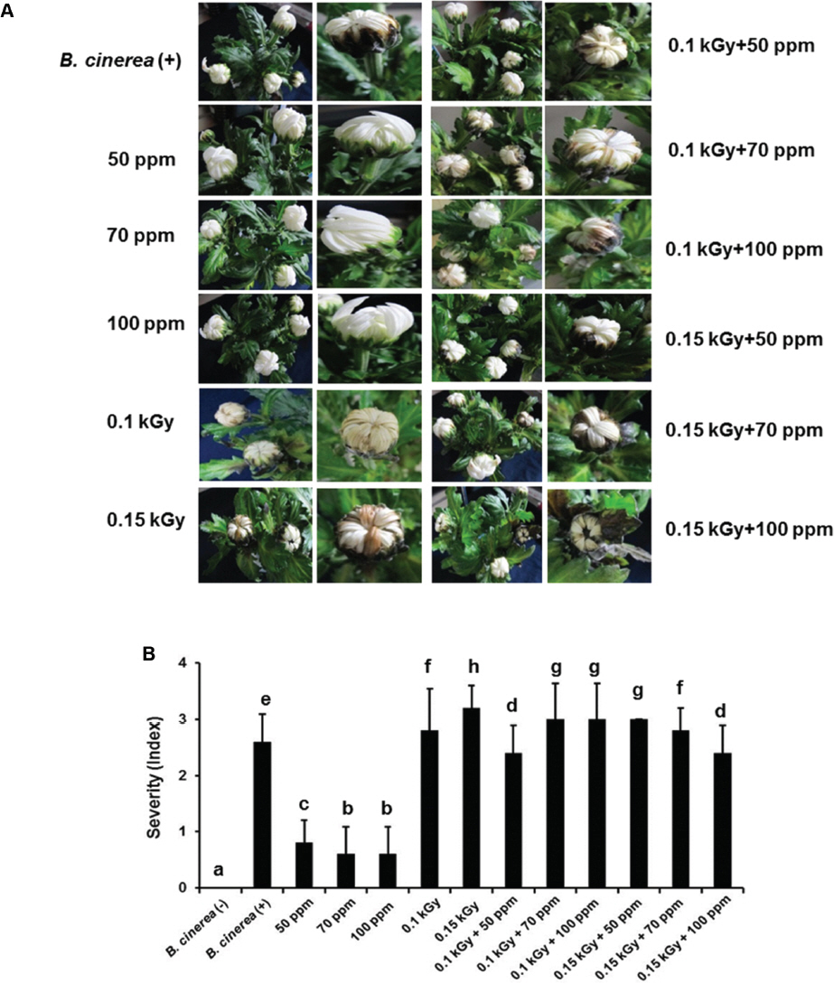
Based on physical properties to gamma irradiation, 0.2 kGy affects the quality of cut chrysanthemum (Fig. 3). Thus, 0.1 or 0.15 kGy gamma irradiation and various concentrations of NaDCC, 50, 70, and 100 ppm, respectively, were used to inhibit the fungal development. The concentration of NaDCC was used less than 100 ppm, since more than 100 ppm of NaDCC causes severe chlorine smell. The chlorine odor was caused in more than 100 ppm of NaDCC, thus NaDCC was used less than 100 ppm of concentrations on NaDCC. Interestingly, as a result of artificial inoculation, the combined treatment with 0.1 or 0.15 kGy of irradiation and 50, 70, and 100 ppm of NaDCC did not inhibit the fungal development in ‘Baekma’. However, single treatment of 50, 70, and 100 ppm of NaDCC significantly inhibited the disease development (Fig. 4A). To monitor the degree of disease index by the treatment, a disease severity index (DSI) (0 to 4) was observed for cut chrysanthemum based on the amount of leaf area infected (Fig. 4B). The combined treatment with 0.1 or 0.15 kGy of irradiation and 50, 70, and 100 ppm of NaDCC did not show reduction in disease severity compared to control. However, single treatment of 50, 70, 100 ppm of NaDCC showed a greater reduction of disease severity compared to the combined treatment. In this study, 50, 70, and 100 ppm of NaDCC, without gamma irradiation, effectively controlled the B. cinerea development on chrysanthemum.
Fig. 4
Effect of gamma irradiation and NaDCC on B. cinerea of cut chrysanthemum flowers. (A) Inhibition of the growth of B. cinerea with gamma irradiation and NaDCC treatment on cut chrysanthemum, ‘Baekma’ flowers. The visual appearance of cut chrysanthemum flowers treated indicated the conditions, and the flowers were photographed at 7 days post treatment. (B) Disease severity in cut chrysanthemum flowers after treatments. The disease severity was scored using a scale of 0 to 4 (0: no infection; 1: 1 to 10% leaf area infected; 2: 11 to 25% leaf area infected; 3: 25 to 50% leaf area infected; 4: 50 to 100% leaf area infected). Values followed by the different letters are significantly different (P<0.05) based on Duncan’s multiple test (n=3).
RPD_21_193_fig_4.jpg

Discussion

Ionizing irradiation is a promising phytosanitary treatment that passes through cut flowers to inactivate harmful pathogens. Many studies have investigated the effects of irradiation on fungi for control of postharvest diseases (Aquino et al., Aziz, 2005 et al., 2007). In this study, gamma irradiation significantly reduced the viability of fungal spores. The spore germination was completely inhibited at 4.0 kGy for B. cinerea, which was considered the lethal dose. However, instead of gamma irradiation, B. cinerea spores exposed electron-beam showed different germination rates, which indicating that the differences in the results may be caused by the types of irradiation source (Chang et al., 1997). Moreover, the same fungal spores showed differences between the two irradiations, gamma irradiation and electron beam, with regards to microbial lethality (Blank and Corrigan, 1995). Thus, it is possible that different physiological mechanisms underlie these response reactions. Although a recent study shows similar effects of gamma irradiation on B. cinerea, the fungal pathogen was isolated from cut roses, not from chrysanthemum (Jeong et al., 2015). It is possible that a fungal pathogen may change their pathogenicity in different hosts. Moreover, the B. cinerea used in this study took from different KACC stock that used in previous study. Thus, these fungal pathogens may show different responses to gamma irradiation.
Single treatment of gamma irradiation is not able to inactivate most microorganisms because of their different radiosensitivities. The cut chrysanthemum will have a negative effect in physio-chemical properties or other quality parameters at doses that required to inactive completely postharvest diseases (Chang et al., 1997). Water uptake and flower opening of irradiated-chrysanthemum did not show a big difference compared to non-irradiated flowers. However, fresh weight was abruptly decreased after 5 days at a dose of over 0.2 kGy of gamma irradiation.
Gamma irradiation was used with alternative agents to increase the efficacy of inhibition of fungal development. Several studies recently suggested that combined treatment with gamma irradiation and eco-friendly agents effectively controlled the postharvest diseases on fruits (Jung et al., 2014; Yoon et al., 2014). NaDCC, also known as sodium dichloro-s-triazinetrione, a chlorine donor, has been approved by the United States Environmental Protection Agency and the World Health Organization for the routine treatment of foods. Nicholl and Prendergast (1998) suggested that 30 min in NaDCC reduced the initial microbial load in various vegetables by 1.69-2.42 log compared to water washed controls. Although NaDCC treatment alone has an effective antibiotic activity, continuous efficiency is not entirely maintained during storage. Thus, a combined treatment of NaDCC and supportive treatment give rise to an effective approach to disinfect pathogens. A previous study showed that an integration of gamma irradiation and NaDCC treatment was effective for satisfactory control of established infections of B. cinerea on paprika (Yoon et al., 2014). However, the combined treatment of gamma irradiation and chlorination did not affect the inhibition of B. cinerea disease development on chrysanthemum. Instead of combined treatment with gamma irradiation, a single treatment of chlorination effectively controlled the B. cinerea development on cut chrysanthemum. This gives rise to the aspect that the activity of inhibition of pathogen development with the combined treatment is associated with postharvest host-pathogen interaction, since B. cinerea of cut chrysanthemum flowers were effectively controlled by the combined treatment (Unpublished data). It is worth emphasizing that researches will deal with the understanding of interactions of a postharvest pathogen with different hosts regarding control by gamma irradiation. Applying NaDCC prior to commercial shipment will be an effective approach to the control of postharvest diseases for cut chrysanthemum flowers. However, single treatment of chlorination may lead to the occurrence of NaDCC-resistant postharvest pathogens (McDonnell and Russell, 1999). Thus, it is necessary to develop an integrated disease control strategy that includes NaDCC. Further investigation is required to evaluate scaling-up to commercial application for the control of other pathogens. Considering these results, it suggests that if the challenges to chlorination treatment are well handled, it will be the ideal approach for the control of postharvest diseases of cut chrysanthemum, and will offer the possibility of accessing a high export value for the market in the future.

Acknowledgements

This research was supported by Export Promotion Technology Development Program, Ministry of Agriculture, Food and Rural Affairs (Project no. 312045-03-3-SB020).

References

Aquino, S, Ferreira, F, Ribeiro, D. H. B, Correa, B, Greiner, R and Villavicencio, A. L. C. H Evaluation of viability of Aspergillus flavus and aflatoxins degradation in irradiated samples of maize. Braz. J. Microbiol 2005. 36: 352-356.
crossref
Aziz, N. H, EI-Far, F. M, Shahin, A. A. M and Roushy, S. M Control of Fusarium moulds and fumonisin B1 in seeds by gamma-irradiation. Food Control 2007. 18: 1337-1342.
crossref
Blank, G and Corrigon, D Comparison of resistance of fungal spore to gamma and electron beam radiation. Int. J. Food Microbiol 1995. 26: 269-277.
crossref pmid
Bradford, M. M A refined and sensitive method for the quantitative of microgram quantities of protein utilizing the principle of protein dye binding. Anal. Biochem 1976. 72: 248-253.
pmid
Bustos-Griffin, E, Hallman, G.J and Griffin, R.L Current and potential trade in horticultural products in irradiated for phytosanitary purposes. Radiat. Phys. Chem 2012. 81: 1203-1207.
crossref
Chang, A. Y, Gladon, R. J, Gleason, M. L, Parker, S. K, Agnew, N. H and Olson, D. G Postharvest quality of cut roses following electron-beam irradiation. HortScience 1997. 32: 698-701.
crossref
De Capdeville, G, Maffia, L. A, Finger, F. L and Batista, U. G Pre-harvest calcium sulfate applications affect vase life and severity of gray mold in cut roses. Sci. Hort 2005. 103: 329-338.
crossref
Dubosis, M, Qilles, K. A, Hamilton, J. K, Rebers, P. A and Smith, F Chemical analysis of microbial cells. Anal. Chem 1956. 28: 350-356.
crossref
Dychdala, G. R ed. by S. S. Block, Chlorine and chlorine compounds. In: Disinfection, Sterilization, and Preservation, 1983. Philadelphia, USA. Lippincott Williams and Wilkins, pp. 157-182.
Elad, Y Latent infection of Botrytis cinerea in rose flowers and combined chemical and physiological control of the disease. Crop Prot 1998. 7: 631-633.
crossref
Fjeld, T, Gislerod, H. R, Revhaug, V and Mortensen, L. M Keeping quality of cut roses as affected by high supplementary irradiation. Sci. Hort 1994. 57: 157-164.
crossref
Geweely, N. S. I and Nawar, L. S Sensitivity to gamma irradiation of post-harvest pathogens of pear. Int. J. Agr. Biol 2006. 6: 710-716.
Hallman, G. J Phytosanitary applications of irradiation. Compr. Rev. Food Sci. Food Saf 2011. 10: 143-151.
crossref
Hammer, P. E, Yang, S. F, Reid, M. S and Marois, J. J Postharvest control of Botrytis cinerea infections on cut roses using fungistatic storage atmospheres. J. Am. Soc. Hort. Sci 1990. 115: 102-107.
crossref
Jeong, R. D, Shin, E. J, Chu, E. H and Park, H. J Effects of ionizing radiation on postharvest fungal pathogens. Plant Pathol. J 2015. 31: 176-180.
crossref pmid pmc
Jung, K, Yoon, M, Park, H. J, Lee, K. Y, Jeong, R. D, Song, B. S and Lee, J. W Application of combined treatment for control of Botrytis cinerea in phytosanitary irradiation processing. Radiat. Phys. Chem 2014. 99: 12-17.
crossref
Lewis, J. A and Papavizas, G. C Permeability changes in hyphae of Rhizoctonia solani induced germling preparations of Trichoderma and Gliocladium. Phytopathology 1987. 77: 699-703.
crossref
McDonnell, G and Russell, A. D Antiseptics and disinfectants: activity, action and, resistance. Clin. Microbiol. Rev 1999. 12: 147-179.
crossref pmid pmc
Nicholl, P and Prendergast, M Disinfection of shredded salad ingredients with sodium dichloroisocyanurate. J. Food Process. Pres 1998. 22: 67-79.
crossref
Rattanawisalanon, C, Ketsa, S. and van Doorn, W. G Effect of aminooxyacetic and sugars on the vase life of Dendrobium flowers. Postharvest Biol. Technol 2003. 29: 93-100.
crossref
Saleh, Y. G, Mayo, M. S and Ahearn, D. G Resistance of some common fungi to gamma irradiation. Appl. Environ. Microbiol 1988. 54: 2134-2135.
crossref pmid pmc
Schwinn, F. J Significance of fungal pathogens in crop production. Pest Outlook 1992. 3: 18-28.
Silva, J. A Chrysanthemum: advances in tissue culture, cryopreservation, postharvest technology, genetic and transgenic biotechnology. Afr. J. Biotechnol 2003. 12: 683-695.
Suslow, T Postharvest Chlorination, Basic Properties and Key Points for Effective Disinfection. Publication 8003. 1997. Oakland, CA, USA. University of California,
Vrind, T. A The Botrytis problem in figures. Acta Hortic 2005. 669: 99-102.
crossref
Williamson, B, Duncan, G. H, Harrison, J. G, Harding, L. A, Elad, Y and Zimand, G Effect of humidity on infecting of rose petals by dry-inoculated conidia of Botrytis cinerea. Mycol. Res 1995. 99: 1303-1310.
Williamson, B, Tudzynski, B, Tudzynski, P and Van Kan, I Botrytis cinerea: the cause of grey mould disease. Mol. Plant Pathol 2007. 8: 561-580.
crossref pmid
Yoon, M, Jung, K, Lee, K. Y, Jeong, J. Y, Lee, J. W and Park, H. J Synergistic effect of the combined treatment with gamma irradiation and sodium dichloroisocyanurate to control gray mold (Botrytis cinerea) on paprika. Radiat. Phys. Chem 2014. 98: 103-108.
crossref


ABOUT
BROWSE ARTICLES
EDITORIAL POLICY
FOR CONTRIBUTORS
Editorial Office
Rm,904 (New Bldg.) The Korean Science & Technology Center 22, Teheran-ro 7-gil, Gangnam-gu, Seoul 06130, Korea
Tel: +82-2-557-9360    Fax: +82-2-557-9361    E-mail: paper@kspp.org                

Copyright © 2024 by The Korean Society of Plant Pathology.

Developed in M2PI

Close layer
prev next