

서론
해바라기(Helianthus annuus L.)는 국화과(Asteraceae)의 1년생 초본식물로 북아메리카 중서부· 지역이 원산지이다(Heiser, 1951). 땅콩, 콩, 유채 등과 더불어 4대 유지작물 중의 하나로 해바라기씨에는 지방유, 필수아미노산이 풍부하여 혈액순환을 도와주며 비타민 함량이 많아 영양학적으로 우수한 식품으로 권장된다(Baudet과 Mossé, 1977; Škorić 등, 2008; Warner, 2005).
세계적으로 해바라기에서 발병되는 주요 병원으로는 풋마름병(Ralstonia solanacearum), 검은무늬병(Alternaria spp.), 잿빛곰팡이병(Botrytis cinerea), 노균병(Plasmopara spp.), 녹병(Puccinia helianthi), 균핵병(Sclerotinia sclerotiorum), 잎마름병(Septoria helianthi), 흰가루병(Sphaerotheca fusca) 등이 있다(Farr과 Rossman, 2016).
해바라기 재배포장에서 해바라기 꽃 부분이 감염이 되어 화뢰(head) 부분이 갈변되었으며 줄기까지 퍼져서 검게 변하였다. 병든 화뢰의 종자로부터 병원균을 순수분리하여 균주를 분리하였으며, 해바라기에 인공접종하여 병원성을 확인하였다. 따라서 본 연구는 해바라기에서 종자의 썩음병을 일으키는 병의 병징, 균학적 특징, 병원성 검정 및 염기서열 분석결과를 보고하고자 한다.
발병 및 병징
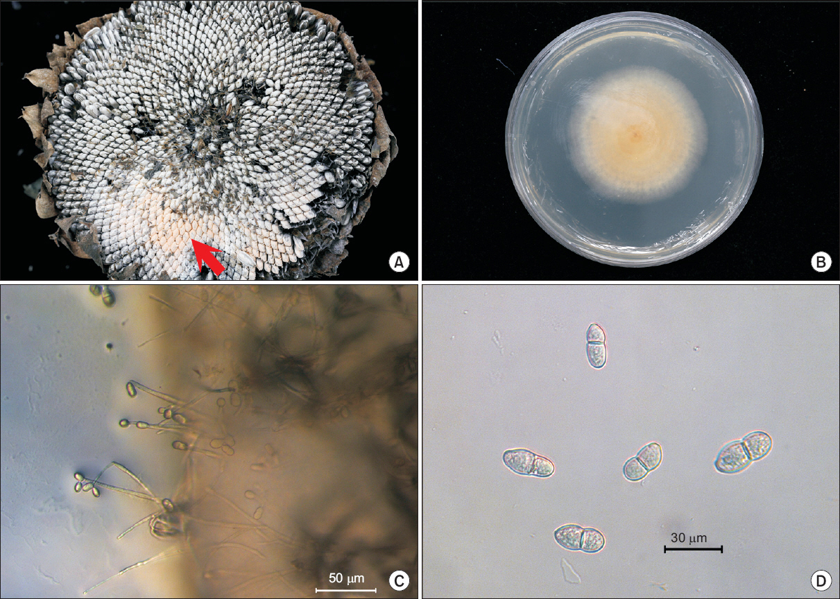
2015년 10월 농업유전자원센터 해바라기 재배포장에서 해바라기 꽃 부분이 검게 변하여 화뢰 부분이 감염되고 줄기까지 검게 변하였다. 종자의 경우는 성숙하지 못하였고 심한 경우 종자가 균사에 덮였으며, 분홍색의 포자층이 형성되기도 했다(Fig. 1A). 병이 진전됨에 따라 줄기가 검게 변하여 고사하였으며, 종자는 여물지 못하고 쭉정이로 남거나 탈락하였다. 이러한 증상은 포장 내에서 여러 곳에 산발적으로 나타났다.
Fig. 1
Symptoms of pink rot on sunflower and microscopic investigation of causal organism. (A) Arrow indicates fungal mass in sunflower head. (B) Colony of isolated fungus on potato dextrose agar (PDA) media at 6 days after inoculation at 25°C. (C) Conidiophores and spores isolated fungus on PDA media. (D) Spores of isolated fungus on PDA media.
병원균의 분리
포장에서 병든 식물체를 수집하여 병원균을 순수분리 배양한 후 균학적 특성과 병원성 검정을 하였다. 병원균을 분리하기 위해 감염된 화뢰 부분의 종자를 70% 에탄올과 1% NaOCl 용액에서 30초간 표면살균하고 멸균수로 2회 세척 후 멸균된 여과지에서 물기를 제거 후 물한천배지(water agar)에 치상하였다. 25°C 항온기에서 3일간 배양한 뒤 균사체 선단부를 떼어 감자한천배지(Difco, Sparks, MD, USA)에 옮겨 25°C에서 배양하였다. 배지에서 형성된 포자로부터 단포자분리를 수행한 뒤 배양하여 포자를 형성시켰다. 균주보관과 병원성 검정을 위해 형성된 포자를 수거하여 20% glycerol에 넣고 -70°C 초저온저장고에 보관하였으며, 병원성 검정 및 염기서열 분석에 사용하였다.
병원균의 균학적 특성
분리된 병원균은 감자한천배지에서 흰색의 균총을 형성하며 자랐으며, 이후에는 주황색의 원형 콜로니를 형성하였으며 동일한 색깔의 분생포자덩어리를 관찰할 수 있었다(Fig. 1B). Conidiophores는 92.9 μm (62.5-123.1 μm)로 단순형 또는 분지형이었다(Fig. 1C). 분생포자는 무색의 격막이 없거나 하나 있는 둥근타원형 혹은 서양배 모양으로 크기는 10.2-21.4-7.5-12.6 μm (평균 16.3-9.7 탆)였다(Fig. 1D). 해바라기에서 분리한 병원균 S068은 기존에 알려진 Trichothecium roseum과 형태적으로 유사하였다(Table 1) (Oh 등, 2014).
Table 1
Morphological characteristics of Trichothecium roseum isolated from sunflower and other hosts
| Structure | Character | S068 | T. roseum* |
|---|---|---|---|
| Colony | Color | Pale roseae | Pale roseae |
| Conidiophores | Shape | Simple or branched below | Simple or branched below |
| Length (μm) | 62.5-123.1 | 150-260 | |
| Conidia | Shape | Ellipsoidal, 2 cell | Ellipsoidal, 2 cell |
| Diameter (μm) | 10.2-21.4×7.5-12.6 | 18-22×8-10 |
* Described by Oh et al. (2014).
병원성 검정
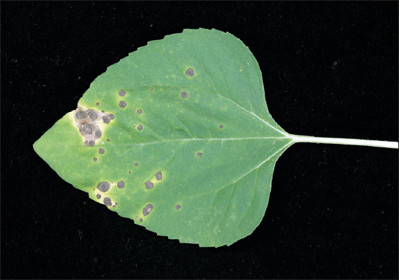
병원성 검정은 파종 후 3주된 유묘에서 실시하였다. 분리된 곰팡이균을 감자한천배지에서 7일간 배양하여 분생포자를 1×10⁶ conidia/ml 농도로 맞추어 건전한 해바라기 유묘에 10 ml씩 분무기로 살포하였으며, 하루 동안 고습도생장상에서 최고 습도를 유지한 뒤 그 후 온실에서 20°C-25°C 온도를 유지하였다. 접종 9일 후 잎에서 검은색의 윤문형 반점이 나타났으며(Fig. 2), 접종한 균주와 동일한 균이 재분리되었다. 따라서 분리한 병원균은 원인병원균으로 판단되었으며 대조구에서는 병징이 나타나지 않았다.
염기서열 분석
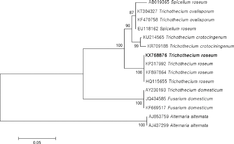
형태적인 특성을 뒷받침하기 위해 염기서열 분석을 실시하였다. 분리한 병원균을 감자한천배지에서 1주일간 배양 후 ribosomal DNA (rDNA)의 internal transcribed spacer (ITS) 영역의 염기서열을 분석하였다(White 등, 1990). Genomic DNA는 DNeasy Plant Mini Kit (Qiagen, Hilden, Germany)를 이용하여 분리하였으며, ITS1/ITS4 primer를 이용하여 PCR 증폭하였다(White 등, 1990). 증폭 산물은 염기서열 분석을 하여 GenBank database (http://blast.ncbi.nlm.nih.gov/Blast.cgi)의 데이터베이스를 이용하여 확인하였다. 계통수 분석은 GenBank 데이터베이스의 ITS 염기서열들을 이용하여 MEGA 6.0 프로그램을 통해 neighbor-joining 방법으로 phylogenetic 분석을 수행하였다. Sequence distance는 Tajima-Nei parameter model로 계산하였다(Saitou와 Nei, 1987; Tamura 등, 2013). 해바라기에서 분리한 병원균 S068의 ITS 염기서열의 크기는 582 bp로 GenBank에 기탁하였다(accession no. KX768876). National Center for Biotechnology Information의 BLAST search 결과 Trichothecium roseum로 등록된 GenBank accession nos. KP317992, KF897865, JX997437 등과 100% 일치하였다. 계통수작성 결과 KX768876의 ITS 염기서열이 T. roseum과 같은 계통군에 속함을 확인할 수 있었다(Fig. 3).
Fig. 3
Phylogenetic analysis of sequence of the internal transcribed spacer ribosomal DNA (rDNA) region of the Trichothecium roseum with closely related strains retrieved from GenBank. The tree was constructed based on the neighbor-joining method with 1,000 replicates. The numbers above the branches represent the bootstrap value. The fungus identified in this study is boldfaced.
T. roseum에 의한 해바라기 수과에서의 병은 조지아, 폴란드에서 보고되어 있으나(Farr과 Rossman, 2016) 우리나라에는 아직 보고되지 않았다(The Korean Society of Plant Pathology, 2009). 따라서 해바라기 화뢰에서 썩음병을 일으키는 균을 분리하여 균학적 특성, 병원성 검정, ITS rDNA 염기서열 비교분석 등의 결과를 바탕으로 T. roseum에 의한 해바라기 분홍빛썩음병이라 명명하고자 한다.
요약
해바라기 재배포장에서 화뢰 부분이 썩는 증상이 나타났다. 병징은 감염된 화뢰 부분이 갈변되어 줄기로 번져갔다. 감염된 화뢰의 종자에는 주황색의 포자덩어리를 관찰할 수 있었다. 병징으로부터 곰팡이를 순수 분리하여 감자한천배지에 배양한 결과, conidiophores에 포자를 형성하여 흰색에서 분홍빛을 띠었다. Conidiophores는 단순형 또는 분지형으로 길이는 62.5-123.1 탆였다. 분생포자는 무색의 격막이 없거나 하나 있는 둥근타원형에서 서양배 모양으로 크기는 10.2-21.4-7.5-12.6 탆였다. 이 균은 건전한 해바라기 잎에 접종하였을 때 잎에서 윤문형의 검은색 반점을 형성하였다. 균학적 특징, 병원성 검정, ITS 염기서열 분석 등의 결과를 바탕으로 Trichothecium roseum으로 동정되었으며 해바라기 분홍빛썩음병으로 명명하고자 한다.
 PDF Links
 PubReader
 Full text via DOI
 Download Citation
 Print
Print


